Laser stimulation of muscle activity with simultaneous detection using a diamond colour centre biosensor
Publication
Metrics
AI Quick Summary
This paper demonstrates the use of a diamond color center biosensor for noninvasively recording muscle activity stimulated by laser optogenetics, capturing both action potentials and slow signal components. The sensor offers a promising alternative to invasive methods, enabling precise, artifact-free monitoring of localized neuromuscular responses.
Paper Preview
Abstract
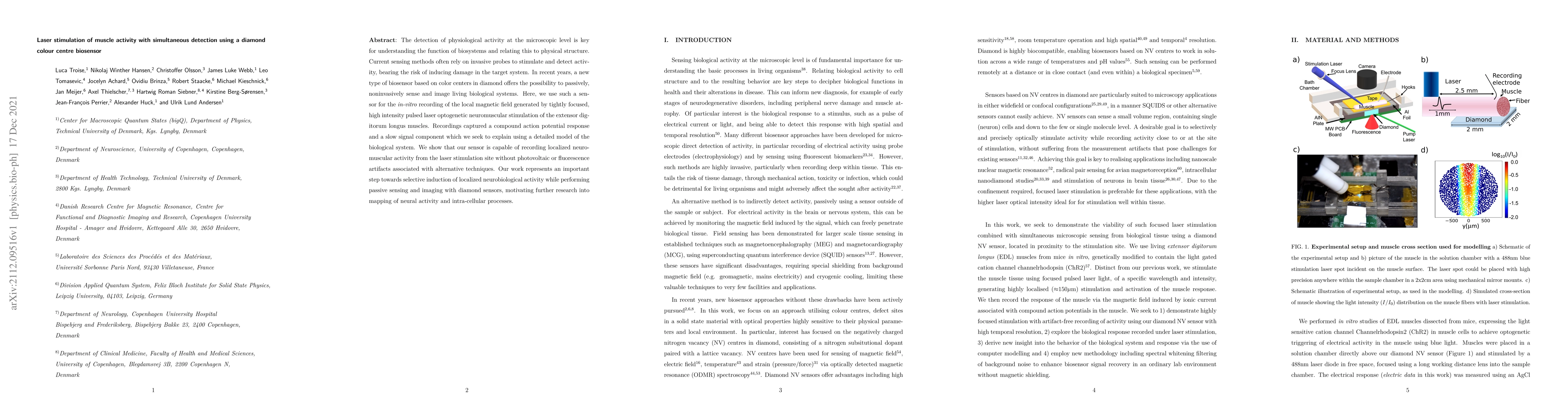
The detection of physiological activity at the microscopic level is key for understanding the function of biosystems and relating this to physical structure. Current sensing methods often rely on invasive probes to stimulate and detect activity, bearing the risk of inducing damage in the target system. In recent years, a new type of biosensor based on color centers in diamond offers the possibility to passively, noninvasively sense and image living biological systems. Here, we use such a sensor for the \textit{in-vitro} recording of the local magnetic field generated by tightly focused, high intensity pulsed laser optogenetic neuromuscular stimulation of the extensor digitorum longus muscles. Recordings captured a compound action potential response and a slow signal component which we seek to explain using a detailed model of the biological system. We show that our sensor is capable of recording localized neuromuscular activity from the laser stimulation site without photovoltaic or fluorescence artifacts associated with alternative techniques. Our work represents an important step towards selective induction of localized neurobiological activity while performing passive sensing and imaging with diamond sensors, motivating further research into mapping of neural activity and intra-cellular processes.
AI Key Findings
Get AI-generated insights about this paper's methodology, results, significance, and more — seven facets brought into focus.
Impact
Paper Details
Authors
PDF Preview
Key Terms
Citation Network
Current paper (gray), citations (green), references (blue)
Display is limited for performance on very large graphs.
Discussion 0