Authors
Summary
Medical images are often high-resolution and lose important detail if downsampled, making pixel-level methods such as semantic segmentation much less efficient if performed on a low-dimensional image. We propose a low-rank Matryoshka projection and a hybrid segmenting architecture that preserves important information while retaining sufficient pixel geometry for pixel-level tasks. We design the Matryoshka Autoencoder (MatAE-U-Net) which combines the hierarchical encoding of the Matryoshka Autoencoder with the spatial reconstruction capabilities of a U-Net decoder, leveraging multi-scale feature extraction and skip connections to enhance accuracy and generalisation. We apply it to the problem of segmenting the left ventricle (LV) in echocardiographic images using the Stanford EchoNet-D dataset, including 1,000 standardised video-mask pairs of cardiac ultrasound videos resized to 112x112 pixels. The MatAE-UNet model achieves a Mean IoU of 77.68\%, Mean Pixel Accuracy of 97.46\%, and Dice Coefficient of 86.91\%, outperforming the baseline U-Net, which attains a Mean IoU of 74.70\%, Mean Pixel Accuracy of 97.31\%, and Dice Coefficient of 85.20\%. The results highlight the potential of using the U-Net in the recursive Matroshka latent space for imaging problems with low-contrast such as echocardiographic analysis.
AI Key Findings
Generated Jun 11, 2025
Methodology
The research methodology involves training a neural network, specifically the Matryoshka Autoencoder-U-Net (MatAE-U-Net) hybrid model, for segmenting the left ventricle in echocardiographic images using the Stanford EchoNet-D dataset. The dataset consists of 1000 manually annotated images, split into 1000 training and 14 testing images. The model employs the Adam optimizer, binary cross-entropy loss, and is implemented using PyTorch.
Key Results
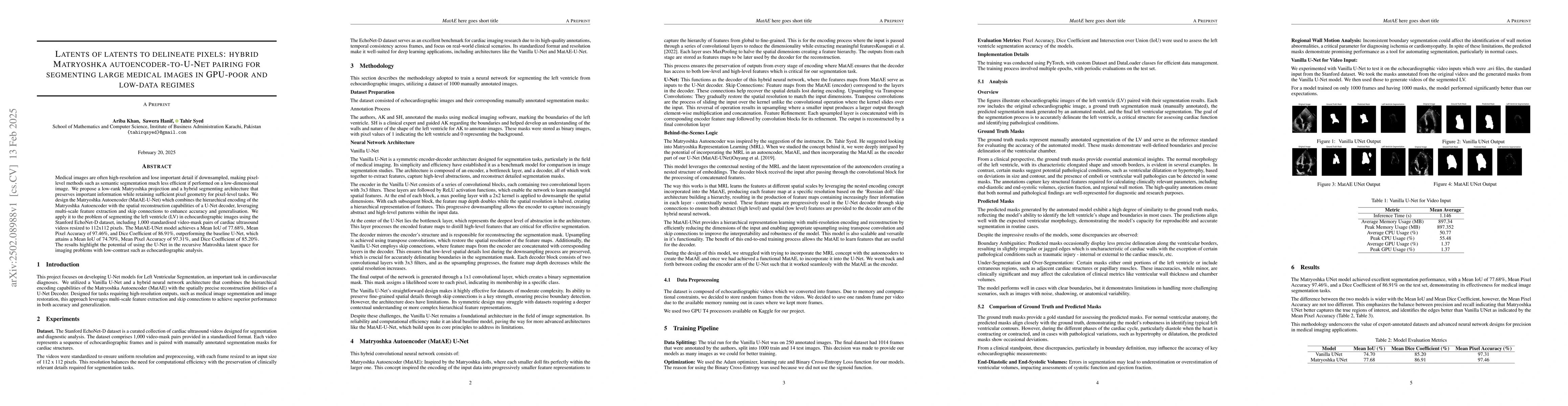
- The MatAE-U-Net model outperforms the baseline Vanilla U-Net model, achieving a Mean IoU of 77.68%, Mean Pixel Accuracy of 97.46%, and Dice Coefficient of 86.91%.
- The MatAE-U-Net model demonstrates improved edge identification and better capture of true regions of interest compared to the Vanilla U-Net, as indicated by the Mean Pixel Accuracy.
Significance
This research is significant as it proposes an efficient solution for segmenting large medical images, specifically in GPU-poor and low-data regimes, which is crucial for tasks like echocardiographic analysis where low-contrast imaging poses challenges.
Technical Contribution
The key technical contribution of this work is the development of the Matryoshka Autoencoder-U-Net (MatAE-U-Net) hybrid model, which combines the hierarchical encoding of the Matryoshka Autoencoder with the spatial reconstruction capabilities of a U-Net decoder, leveraging multi-scale feature extraction and skip connections to enhance accuracy and generalization.
Novelty
This work introduces a novel hybrid architecture, MatAE-U-Net, that effectively addresses the challenges of segmenting high-resolution medical images with limited computational resources and data, by preserving important information and maintaining sufficient pixel geometry for pixel-level tasks.
Limitations
- The study was limited to segmenting the left ventricle in echocardiographic images and did not explore other medical imaging modalities or anatomical structures.
- The model's performance in handling more challenging scenarios, such as images with noise, shadowing, or anatomical variability, was not extensively evaluated.
Future Work
- Further research could explore the application of this model to other medical imaging tasks and datasets.
- Investigating the model's performance in handling more complex and diverse image datasets could enhance its robustness and generalizability.
Paper Details
PDF Preview
Citation Network
Current paper (gray), citations (green), references (blue)
Display is limited for performance on very large graphs.
Similar Papers
Found 4 papersLinkGAN: Linking GAN Latents to Pixels for Controllable Image Synthesis
Qifeng Chen, Jiapeng Zhu, Bo Dai et al.
Segmenting Medical Images with Limited Data
Zhaoshan Liua, Qiujie Lv, Chau Hung Lee et al.
Beyond Input Activations: Identifying Influential Latents by Gradient Sparse Autoencoders
Haiyan Zhao, Dong Shu, Mengnan Du et al.
No citations found for this paper.
Comments (0)