Authors
Summary
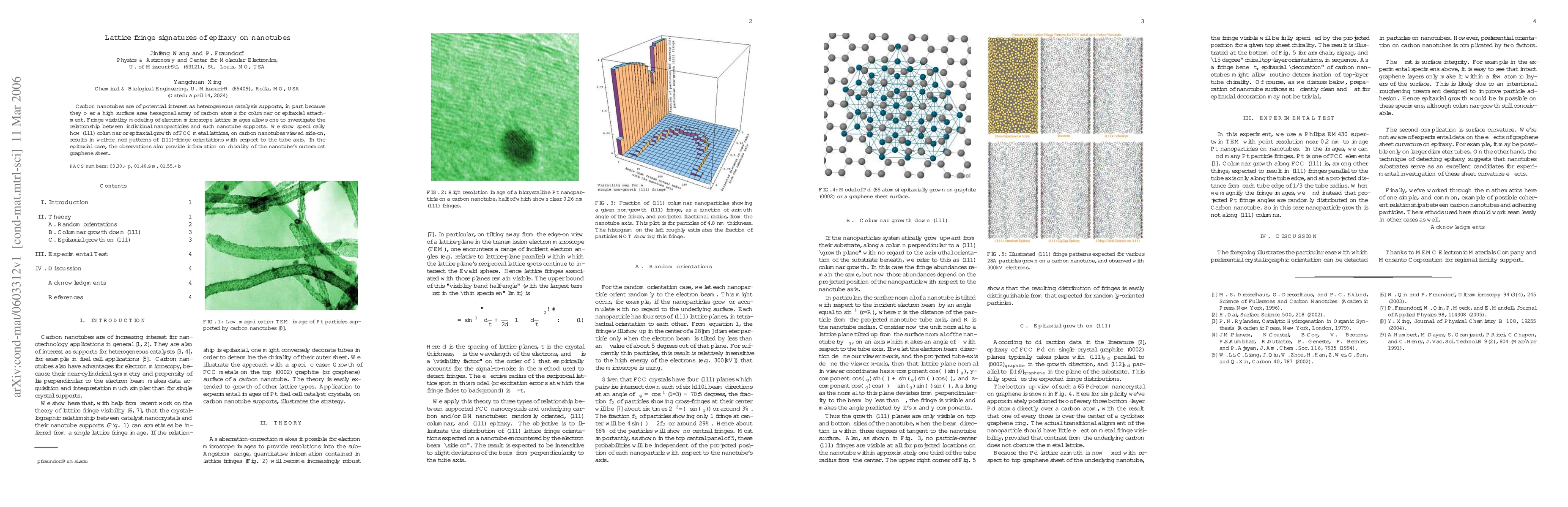
Carbon nanotubes are of potential interest as heterogeneous catalysis supports, in part because they offer a high surface area hexagonal array of carbon atoms for columnar or epitaxial attachment. Fringe visibility modeling of electron microscope lattice images allows one to investigate the relationship between individual nanoparticles and such nanotube supports. We show specifically how (111) columnar or epitaxial growth of FCC metal lattices, on carbon nanotubes viewed side-on, results in well-defined patterns of (111)-fringe orientations with respect to the tube axis. In the epitaxial case, the observations also provide information on chirality of the nanotube's outermost graphene sheet.
AI Key Findings
Get AI-generated insights about this paper's methodology, results, and significance.
Paper Details
PDF Preview
Citation Network
Current paper (gray), citations (green), references (blue)
Display is limited for performance on very large graphs.
Similar Papers
Found 4 papersStranger Danger! Cross-Community Interactions with Fringe Users Increase the Growth of Fringe Communities on Reddit
Robert West, Giuseppe Russo, Manoel Horta Ribeiro
| Title | Authors | Year | Actions |
|---|
Comments (0)