Summary
Smart systems that can accurately diagnose patients with mental disorders and identify effective treatments based on brain functional imaging data are of great applicability and are gaining much attention. Most previous machine learning studies use hand-designed features, such as functional connectivity, which does not maintain the potential useful information in the spatial relationship between brain regions and the temporal profile of the signal in each region. Here we propose a new method based on recurrent-convolutional neural networks to automatically learn useful representations from segments of 4-D fMRI recordings. Our goal is to exploit both spatial and temporal information in the functional MRI movie (at the whole-brain voxel level) for identifying patients with schizophrenia.
AI Key Findings
Generated Sep 02, 2025
Methodology
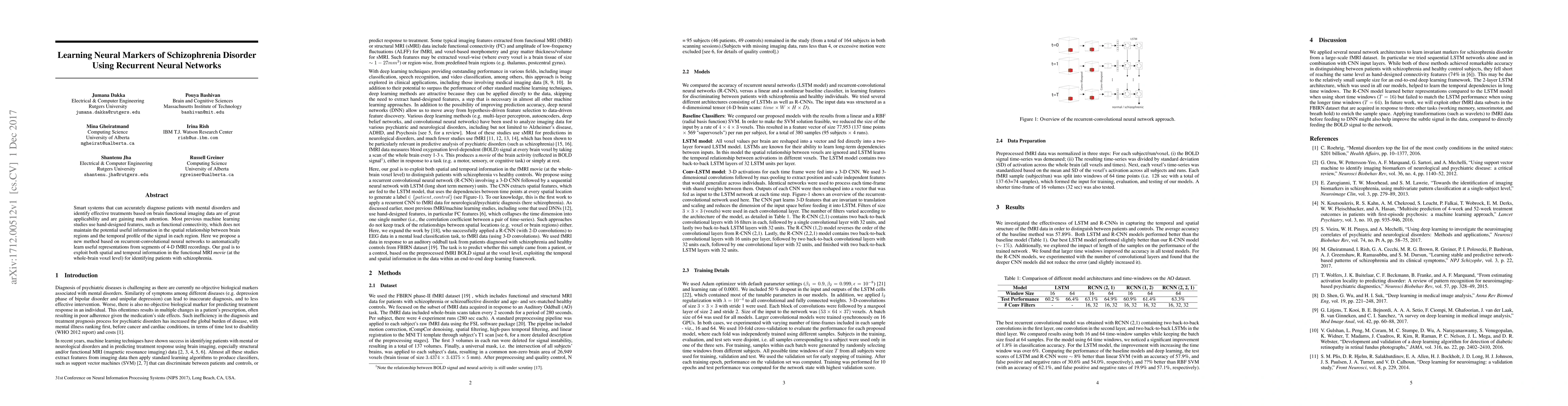
The research proposes a method using recurrent-convolutional neural networks (R-CNN) to automatically learn representations from 4-D fMRI recordings, exploiting both spatial and temporal information for distinguishing patients with schizophrenia from healthy controls.
Key Results
- LSTM and R-CNN models outperformed a baseline linear and RBF SVM, achieving test performance of 60.2% and 66.4% respectively.
- The best R-CNN model (RCNN(2,1)) contained two back-to-back convolutional layers in the first layer, one convolution in the second layer, and two back-to-back LSTM layers in the third layer.
- Larger time windows (64 samples) significantly improved the accuracy in all tested models, with a 1.8% improvement for R-CNN models and over 6% for LSTM models.
Significance
This research is significant as it aims to develop smart systems for accurate diagnosis of mental disorders like schizophrenia and identifying effective treatments using brain functional imaging data, potentially surpassing the performance of traditional machine learning techniques.
Technical Contribution
The paper introduces a novel approach using R-CNNs to directly learn from fMRI data without the need for hand-designed features, allowing for data-driven feature discovery and potentially improving prediction accuracy.
Novelty
This work is novel as it is among the first to apply recurrent CNNs to fMRI data for neurological/psychiatric diagnosis, unlike previous studies that mainly used hand-crafted features or 2D convolutions on EEG data.
Limitations
- The study's sample size might be relatively small for an end-to-end deep learning framework, which could limit the models' performance compared to hand-designed connectivity features.
- The relationship between BOLD signal and neural activity is still under scrutiny.
Future Work
- Explore other fMRI data subsets from the FBIRN dataset acquired in response to different tasks to enrich the sample space.
- Apply transformations, such as wavelets, to fMRI data before feeding it into deep neural networks to potentially improve the detection of subtle signals in the data.
Paper Details
PDF Preview
Key Terms
Citation Network
Current paper (gray), citations (green), references (blue)
Display is limited for performance on very large graphs.
Similar Papers
Found 4 papers| Title | Authors | Year | Actions |
|---|
Comments (0)