Learning to Reconstruct Confocal Microscopy Stacks from Single Light Field Images
Publication
Metrics
AI Quick Summary
This paper introduces a deep learning method using LFMNet to reconstruct 3D confocal microscopy stacks from single light field images, significantly reducing scan time and storage requirements. The approach was evaluated on mouse brain slices, demonstrating its potential for real-time in vivo 3D microscopy.
Paper Preview
Abstract
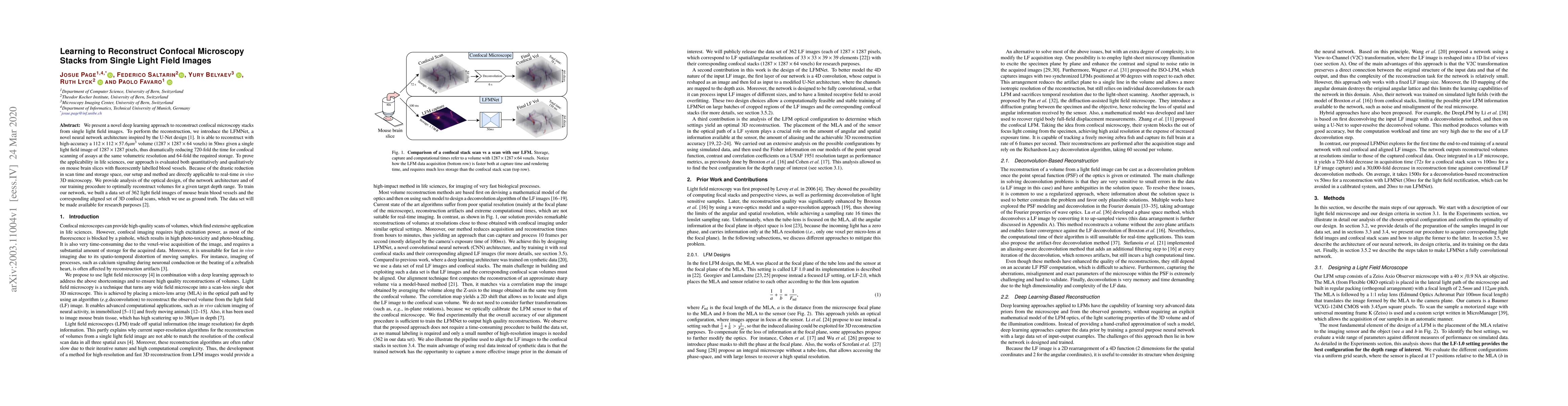
We present a novel deep learning approach to reconstruct confocal microscopy stacks from single light field images. To perform the reconstruction, we introduce the LFMNet, a novel neural network architecture inspired by the U-Net design. It is able to reconstruct with high-accuracy a 112x112x57.6$\mu m^3$ volume (1287x1287x64 voxels) in 50ms given a single light field image of 1287x1287 pixels, thus dramatically reducing 720-fold the time for confocal scanning of assays at the same volumetric resolution and 64-fold the required storage. To prove the applicability in life sciences, our approach is evaluated both quantitatively and qualitatively on mouse brain slices with fluorescently labelled blood vessels. Because of the drastic reduction in scan time and storage space, our setup and method are directly applicable to real-time in vivo 3D microscopy. We provide analysis of the optical design, of the network architecture and of our training procedure to optimally reconstruct volumes for a given target depth range. To train our network, we built a data set of 362 light field images of mouse brain blood vessels and the corresponding aligned set of 3D confocal scans, which we use as ground truth. The data set will be made available for research purposes.
AI Key Findings
Get AI-generated insights about this paper's methodology, results, significance, and more — seven facets brought into focus.
Impact
Paper Details
Authors
PDF Preview
Key Terms
Citation Network
Current paper (gray), citations (green), references (blue)
Display is limited for performance on very large graphs.
Discussion 0