This paper introduces a deep neural network for microscopy image segmentation that can handle coarse labels and minimal pixel-wise annotations, improving efficiency and flexibility in training data-hungry models.
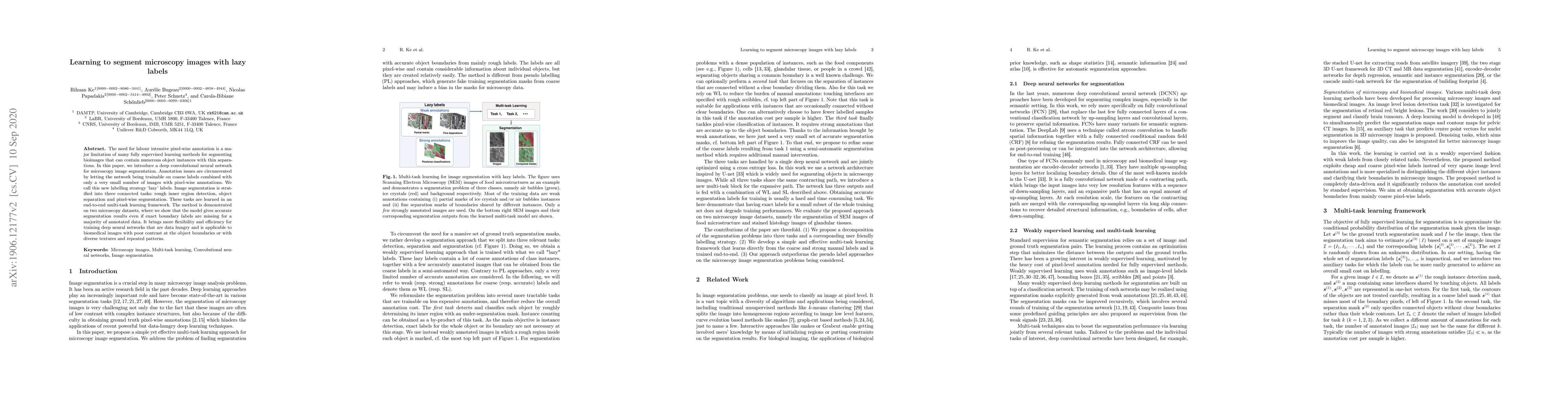
The need for labour intensive pixel-wise annotation is a major limitation of many fully supervised learning methods for segmenting bioimages that can contain numerous object instances with thin separations. In this paper, we introduce a deep convolutional neural network for microscopy image segmentation. Annotation issues are circumvented by letting the network being trainable on coarse labels combined with only a very small number of images with pixel-wise annotations. We call this new labelling strategy `lazy' labels. Image segmentation is stratified into three connected tasks: rough inner region detection, object separation and pixel-wise segmentation. These tasks are learned in an end-to-end multi-task learning framework. The method is demonstrated on two microscopy datasets, where we show that the model gives accurate segmentation results even if exact boundary labels are missing for a majority of annotated data. It brings more flexibility and efficiency for training deep neural networks that are data hungry and is applicable to biomedical images with poor contrast at the object boundaries or with diverse textures and repeated patterns.
Get AI-generated insights about this paper's methodology, results, and significance.
Current paper (gray), citations (green), references (blue)
Qingyuan Liu, Avideh Zakhor
Title | Authors | Year | Actions |
---|---|---|---|
Multi-task Learning Network for CT Whole Heart Segmentation
|
Jianqin Yin, Jin Liu, Junying Wang, Jun Liu
|
2023
|
View Paper |
A State-of-the-art Survey of U-Net in Microscopic Image Analysis: from Simple Usage to Structure Mortification
|
Jian Wu, Wanli Liu, Chen Li, Tao Jiang, Islam Mohammad Shariful, Hongzan Sun, Xiaoqi Li, Xintong Li, Xinyu Huang, M. Grzegorzek
|
2022
|
View Paper |
LR-Net: A Multi-task Model Using Relationship-based Contour Information to Enhance the Semantic Segmentation of Cancer Regions
|
Baorong Shi, Hong Zhang, Rui Yan, Wang Jing, Jinfeng Zang, Fa Zhang
|
2020
|
View Paper |
A Three-Stage Self-Training Framework for Semi-Supervised Semantic Segmentation
|
Rihuan Ke, Angelica I. Avilés-Rivero, Saurabh Pandey, Saikumar Reddy, C. Schönlieb
|
2020
|
View Paper |
Application of Artificial Neural Networks for Analysis of Ice Recrystallization Process for Cryopreservation
|
M. Tymkovych, O. Gryshkov, K. Selivanova, V. Mutsenko, Oleg Avrunin, B. Glasmacher
|
2020
|
View Paper |
An anatomically-informed 3D CNN for brain aneurysm classification with weak labels
|
Tommaso Di Noto, Guillaume Marie, S. Tourbier, Yasser Alem'an-G'omez, G. Saliou, M. Cuadra, P. Hagmann, Jonas Richiardi
|
2020
|
View Paper |
Holistic multitask regression network for multiapplication shape regression segmentation
|
Clara M. Tam, Dong Zhang, Bo Chen, T. Peters, Shuo Li
|
2020
|
View Paper |
Deep semantic segmentation of natural and medical images: a review
|
Saeid Asgari Taghanaki, Kumar Abhishek, Joseph Paul Cohen, J. Cohen-Adad, G. Hamarneh
|
2019
|
View Paper |
Assisting human experts in the interpretation of their visual process: A case study on assessing copper surface adhesive potency
|
T. Hascoet, Xuejiao Deng, Kiyoto Tai, Mari Sugiyama, Yuji Adachi, Sachiko Nakamura, Y. Ariki, Tomoko Hayashi, T. Takiguchi
|
2019
|
View Paper |
Machine Learning towards General Medical Image Segmentation
|
Clara M. Tam
|
2020
|
View Paper |
Comments (0)