Contrast-Enhanced Spectral Mammography (CESM) is a dual-energy mammographic
technique that improves lesion visibility through the administration of an
iodinated contrast agent. It acquires both a low-energy image, comparable to
standard mammography, and a high-energy image, which are then combined to
produce a dual-energy subtracted image highlighting lesion contrast
enhancement. While CESM offers superior diagnostic accuracy compared to
standard mammography, its use entails higher radiation exposure and potential
side effects associated with the contrast medium. To address these limitations,
we propose Seg-CycleGAN, a generative deep learning framework for Virtual
Contrast Enhancement in CESM. The model synthesizes high-fidelity dual-energy
subtracted images from low-energy images, leveraging lesion segmentation maps
to guide the generative process and improve lesion reconstruction. Building
upon the standard CycleGAN architecture, Seg-CycleGAN introduces localized loss
terms focused on lesion areas, enhancing the synthesis of diagnostically
relevant regions. Experiments on the CESM@UCBM dataset demonstrate that
Seg-CycleGAN outperforms the baseline in terms of PSNR and SSIM, while
maintaining competitive MSE and VIF. Qualitative evaluations further confirm
improved lesion fidelity in the generated images. These results suggest that
segmentation-aware generative models offer a viable pathway toward
contrast-free CESM alternatives.
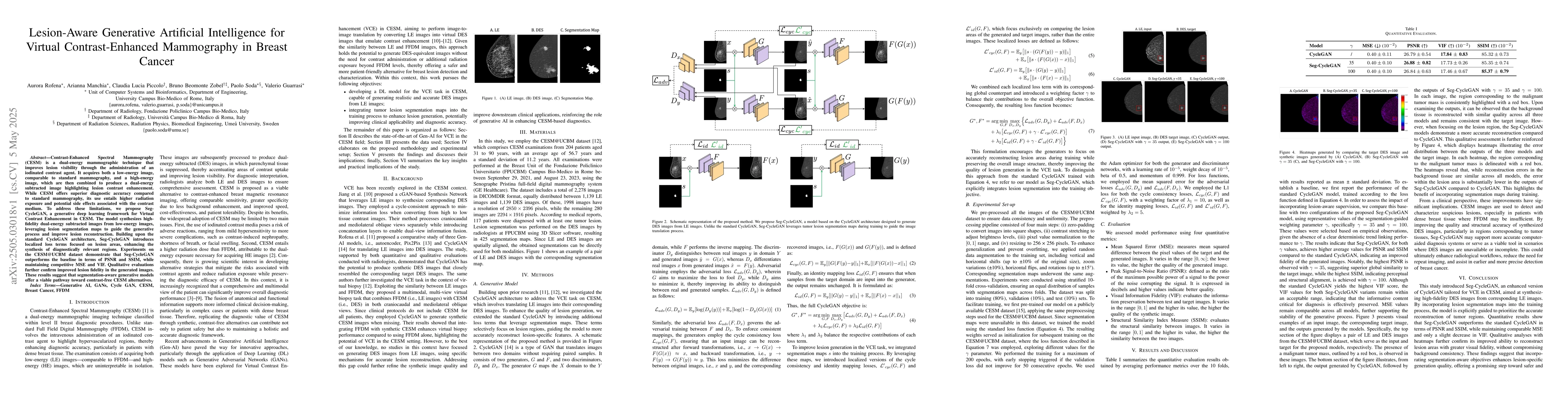
Discussion 0