ArxivLens

Quick Summary:

LesionMix introduces a lesion-level data augmentation method to enhance medical image segmentation by augmenting specific abnormalities within images. Experiments show it outperforms other Mix-based methods, improving lesion shape, location, and intensity diversity across various datasets.

arXiv Id: 2308.09026
Comments: 13 pages, 5 figures, 4 tables, MICCAI DALI Workshop 2023
Date Published: 2023-08-21
Date Updated: 2023-08-21

Categories:

Key Terms (TF-IDF):

augmentation (0.334)
data augmentation (0.325)
image (0.230)
diversity (0.202)
medical (0.197)
segmentation (0.195)
intensity (0.188)
ct (0.151)
medical image (0.148)
datasets (0.145)
outperforming (0.139)
brain (0.132)
load (0.132)
data (0.130)
modalities (0.129)
level (0.128)
released (0.126)
learning based (0.121)
aware (0.120)
performs (0.119)
License: http://creativecommons.org/licenses/by/4.0/

Summary:

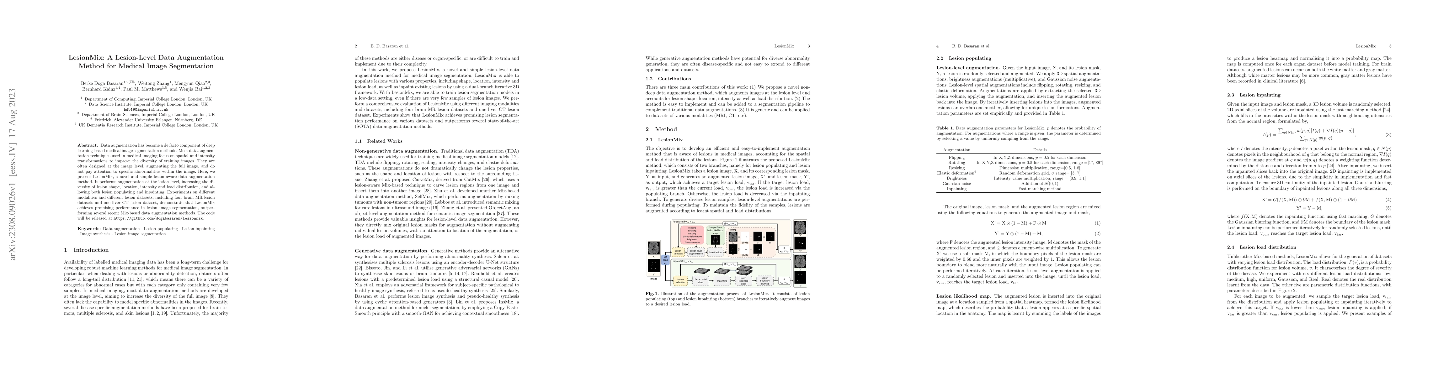
Data augmentation has become a de facto component of deep learning-based medical image segmentation methods. Most data augmentation techniques used in medical imaging focus on spatial and intensity transformations to improve the diversity of training images. They are often designed at the image level, augmenting the full image, and do not pay attention to specific abnormalities within the image. Here, we present LesionMix, a novel and simple lesion-aware data augmentation method. It performs augmentation at the lesion level, increasing the diversity of lesion shape, location, intensity and load distribution, and allowing both lesion populating and inpainting. Experiments on different modalities and different lesion datasets, including four brain MR lesion datasets and one liver CT lesion dataset, demonstrate that LesionMix achieves promising performance in lesion image segmentation, outperforming several recent Mix-based data augmentation methods. The code will be released at https://github.com/dogabasaran/lesionmix.

Citation & Reference Network Visualization

This graph shows how this paper is connected to other research. The current paper is shown in dark gray, papers that cite it are in green, and papers it references are in blue. You can:

Current Paper
Papers citing this work (Citations)
Papers cited by this work (References)

• Hover over any node to see the paper title

• Click a node to view that paper's details

• Drag nodes to rearrange the graph

• Use the zoom controls (bottom right) to zoom in/out

Citations and References

Title Authors Year Actions
SegHeD+: Segmentation of Heterogeneous Data for Multiple Sclerosis Lesions with Anatomical Constraints and Lesion-aware Augmentation
Berke Doga Basaran, Paul M. Matthews, Wenjia Bai
2024
View Paper
SegHeD: Segmentation of Heterogeneous Data for Multiple Sclerosis Lesions with Anatomical Constraints
Berke Doga Basaran, Xinru Zhang, Paul M. Matthews, Wenjia Bai
2024
View Paper
HSMix: Hard and soft mixing data augmentation for medical image segmentation
D. Sun, F. Dornaika, N. Barrena
2024
View Paper
Analyzing Tumors by Synthesis
Qi Chen, Yuxiang Lai, Xiaoxi Chen, Qixing Hu, Alan L. Yuille, Zongwei Zhou
2024
View Paper
A Foundation Model for Brain Lesion Segmentation with Mixture of Modality Experts
Xinru Zhang, N. Ou, Berke Doga Basaran, Marco Visentin, Mengyun Qiao, Renyang Gu, Ouyang Cheng, Yaou Liu, Paul M. Matthew, Chuyang Ye, Wenjia Bai
2024
View Paper
A tumor inpainting framework for MRI using automated masks based on channel-specific conditions across the volume
Kyuri Kim, Hyungjoo Cho, Yoonho Na, Sung-Joon Ye, Jimin Lee, Sungsoo Ahn, Ji Eun Park, Hwiyoung Kim
2025
View Paper

Comments (0)

This website uses cookies to ensure you get the best experience. Learn more