Publication
Metrics
AI Quick Summary
This study employs convolutional neural networks (CNNs) to reconstruct 3D angiographic volumes from sparse cone beam CT projections, validated through patient-specific intracranial aneurysm geometries and computational fluid dynamics simulations. The trained U-Net model achieved a mean squared error of 0.0001, demonstrating the potential of CNNs for enhancing temporal resolution in 3D angiography.
Paper Preview
Abstract
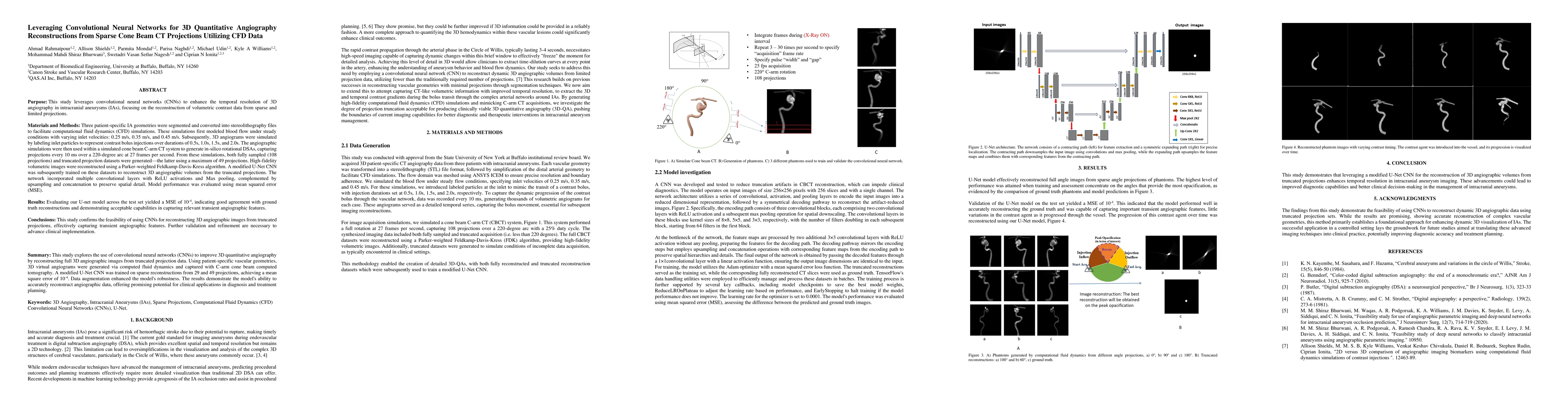
This study leverages convolutional neural networks to enhance the temporal resolution of 3D angiography in intracranial aneurysms focusing on the reconstruction of volumetric contrast data from sparse and limited projections. Three patient-specific IA geometries were segmented and converted into stereolithography files to facilitate computational fluid dynamics simulations. These simulations first modeled blood flow under steady conditions with varying inlet velocities: 0.25 m/s, 0.35 m/s, and 0.45 m/s. Subsequently, 3D angiograms were simulated by labeling inlet particles to represent contrast bolus injections over durations of 0.5s, 1.0s, 1.5s, and 2.0s. The angiographic simulations were then used within a simulated cone beam C arm CT system to generate in-silico rotational DSAs, capturing projections every 10 ms over a 220-degree arc at 27 frames per second. From these simulations, both fully sampled (108 projections) and truncated projection datasets were generated the latter using a maximum of 49 projections. High fidelity volumetric images were reconstructed using a Parker weighted Feldkamp Davis Kress algorithm. A modified U Net CNN was subsequently trained on these datasets to reconstruct 3D angiographic volumes from the truncated projections. The network incorporated multiple convolutional layers with ReLU activations and Max pooling, complemented by upsampling and concatenation to preserve spatial detail. Model performance was evaluated using mean squared error (MSE). Evaluating our U net model across the test set yielded a MSE of 0.0001, indicating good agreement with ground truth reconstructions and demonstrating acceptable capabilities in capturing relevant transient angiographic features. This study confirms the feasibility of using CNNs for reconstructing 3D angiographic images from truncated projections.
AI Key Findings
Get AI-generated insights about this paper's methodology, results, significance, and more — seven facets brought into focus.
Impact
Authors
PDF Preview
Citation Network
Current paper (gray), citations (green), references (blue)
Display is limited for performance on very large graphs.
Discussion 0