Publication
Metrics
AI Quick Summary
This paper presents a method for upconversion microscopy using a 970-nm LED instead of high-power lasers, achieving single-nanoparticle sensitivity despite lower efficiency and higher background. Time-gated luminescence detection mitigates background, enabling clear imaging of UCNPs and demonstrating cellular uptake in cells.
Paper Preview
Abstract
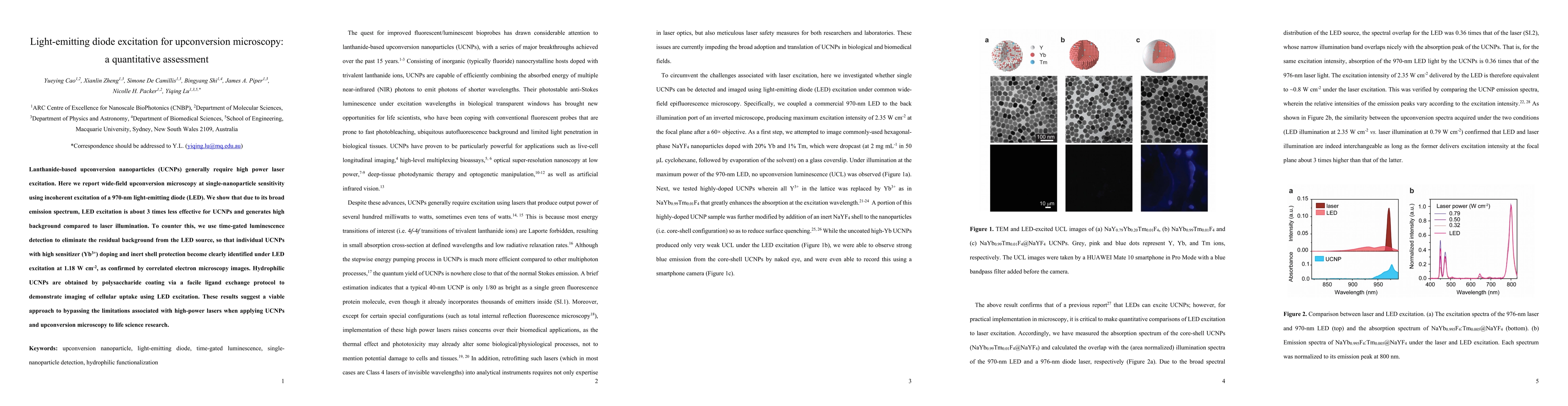
Lanthanide-based upconversion nanoparticles (UCNPs) generally require high power laser excitation. Here we report wide-field upconversion microscopy at single-nanoparticle sensitivity using incoherent excitation of a 970-nm light-emitting diode (LED). We show that due to its broad emission spectrum, LED excitation is about 3 times less effective for UCNPs and generates high background compared to laser illumination. To counter this, we use time-gated luminescence detection to eliminate the residual background from the LED source, so that individual UCNPs with high sensitizer (Yb3+) doping and inert shell protection become clearly identified under LED excitation at 1.18 W cm-2, as confirmed by correlated electron microscopy images. Hydrophilic UCNPs are obtained by polysaccharide coating via a facile ligand exchange protocol to demonstrate imaging of cellular uptake using LED excitation. These results suggest a viable approach to bypassing the limitations associated with high-power lasers when applying UCNPs and upconversion microscopy to life science research.
AI Key Findings
Get AI-generated insights about this paper's methodology, results, significance, and more — seven facets brought into focus.
Impact
Paper Details
Authors
PDF Preview
Key Terms
Citation Network
Current paper (gray), citations (green), references (blue)
Display is limited for performance on very large graphs.
Discussion 0