Publication
Metrics
AI Quick Summary
This paper surveys advanced computational methods for light-field microscopy (LFM) in neuroscience, emphasizing the integration of model-based and data-driven approaches to enhance the interpretability and generalization of imaging neuronal activity. It highlights the potential of combining signal processing and wave-optics theories to develop novel techniques for high-speed 3D imaging.
Paper Preview
Abstract
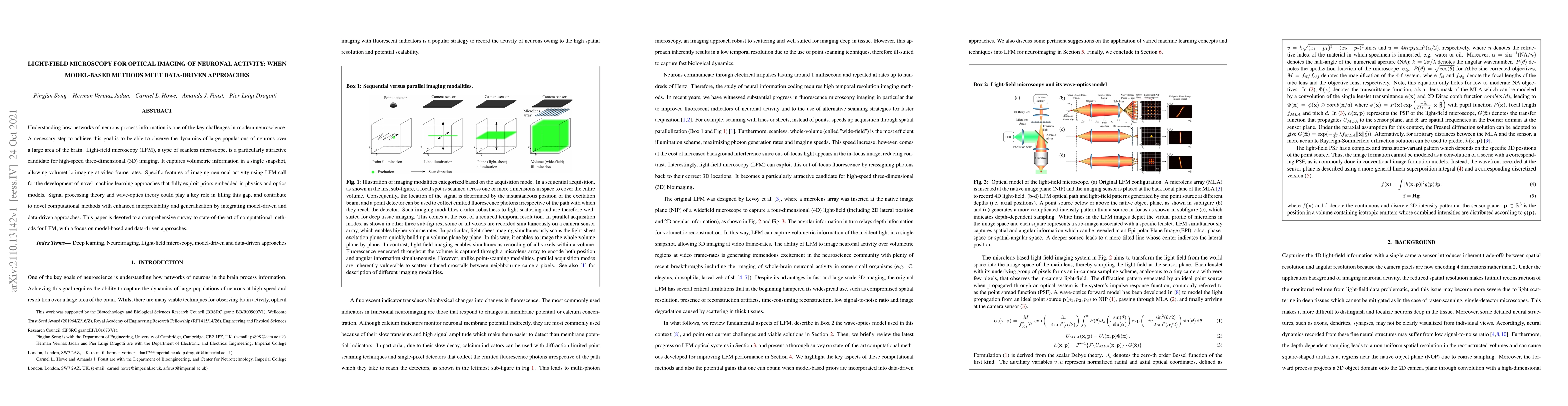
Understanding how networks of neurons process information is one of the key challenges in modern neuroscience. A necessary step to achieve this goal is to be able to observe the dynamics of large populations of neurons over a large area of the brain. Light-field microscopy (LFM), a type of scanless microscope, is a particularly attractive candidate for high-speed three-dimensional (3D) imaging. It captures volumetric information in a single snapshot, allowing volumetric imaging at video frame-rates. Specific features of imaging neuronal activity using LFM call for the development of novel machine learning approaches that fully exploit priors embedded in physics and optics models. Signal processing theory and wave-optics theory could play a key role in filling this gap, and contribute to novel computational methods with enhanced interpretability and generalization by integrating model-driven and data-driven approaches. This paper is devoted to a comprehensive survey to state-of-the-art of computational methods for LFM, with a focus on model-based and data-driven approaches.
AI Key Findings
Get AI-generated insights about this paper's methodology, results, significance, and more — seven facets brought into focus.
Impact
Paper Details
Authors
PDF Preview
Key Terms
Citation Network
Current paper (gray), citations (green), references (blue)
Display is limited for performance on very large graphs.
Discussion 0