Quick Summary:

This paper proposes a deep image prior (DIP) framework for limited-angle SPECT image reconstruction, which incorporates a forward projection model and a binary mask to restore non-collected projection data, significantly improving image quality and reducing distortion. The method outperforms existing back-projection techniques, as demonstrated by higher PSNR and SSIM values in numerical simulations and clinical data.

Summary

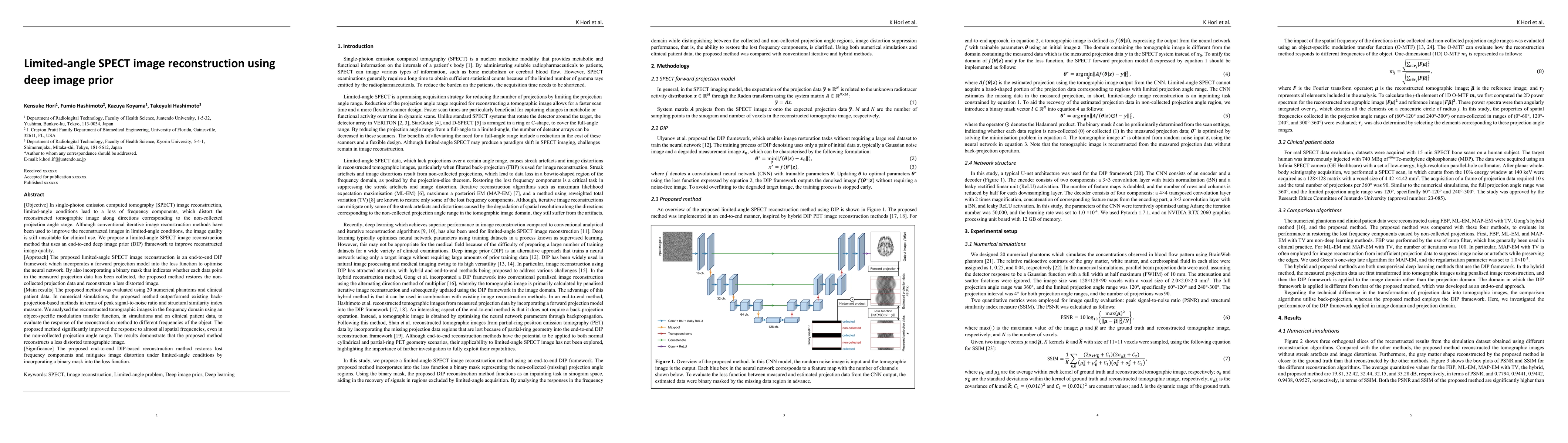
In SPECT image reconstruction, limited-angle (LA) conditions lead to a loss of frequency components, which distort the reconstructed tomographic image along directions corresponding to the non-collected projection angle range. Although conventional iterative image reconstruction methods have been used to improve the reconstructed images in LA conditions, the image quality is still unsuitable for clinical use. We propose a LA SPECT image reconstruction method that uses an end-to-end deep image prior (DIP) framework to improve reconstructed image quality. The proposed LA SPECT image reconstruction is an end-to-end DIP framework which incorporates a forward projection model into the loss function to optimise the neural network. By also incorporating a binary mask that indicates whether each data point in the measured projection data has been collected, the proposed method restores the non-collected projection data and reconstructs a less distorted image. The proposed method was evaluated using 20 numerical phantoms and clinical patient data. In numerical simulations, the proposed method outperformed existing back-projection-based methods in terms of PSNR and SSIM. We analysed the reconstructed tomographic images in the frequency domain using an object-specific modulation transfer function, in simulations and on clinical patient data, to evaluate the response of the reconstruction method to different frequencies of the object. The proposed method significantly improved the response to almost all spatial frequencies, even in the non-collected projection angle range. The results demonstrate that the proposed method reconstructs a less distorted tomographic image. The proposed end-to-end DIP-based reconstruction method restores lost frequency components and mitigates image distortion under LA conditions by incorporating a binary mask into the loss function.

AI Key Findings

Generated Jun 10, 2025

Methodology

The research proposes a limited-angle SPECT image reconstruction method using an end-to-end deep image prior (DIP) framework, which incorporates a forward projection model and a binary mask into the loss function to optimize the neural network and restore non-collected projection data.

Key Results

  • The proposed method outperformed existing back-projection-based methods in terms of PSNR and SSIM in numerical simulations.
  • The proposed method significantly improved the response to almost all spatial frequencies, even in the non-collected projection angle range, as analyzed using an object-specific modulation transfer function (O-MTF) on both simulations and clinical patient data.
  • The proposed method reconstructed tomographic images without streak artifacts and image distortions, as demonstrated in both numerical phantoms and clinical patient data.

Significance

This research is significant as it addresses the issue of image distortion in SPECT imaging under limited-angle conditions, which is crucial for improving image quality suitable for clinical use.

Technical Contribution

The main technical contribution is the end-to-end DIP-based reconstruction method that restores lost frequency components and mitigates image distortion under limited-angle conditions by incorporating a binary mask into the loss function.

Novelty

This work is novel as it introduces a deep learning approach to limited-angle SPECT image reconstruction, leveraging the DIP framework and a forward projection model to address frequency loss and image distortion issues not effectively solved by conventional iterative methods.

Limitations

  • The learning rate was set to a small value (1.0×10^-4) to ensure stable convergence, resulting in long computation times that may not be suitable for clinical applications without optimization.
  • The study did not explore the applicability of the proposed method to various organs or imaging purposes beyond bone SPECT

Future Work

  • Investigate the use of block iterative algorithms or pre-trained neural network parameters to reduce computation time and GPU memory usage for clinical applicability.
  • Expand the application of the proposed method to other organs and imaging purposes to enhance its clinical relevance.

Paper Details

PDF Preview

Download PDF

Key Terms

No key terms available for this paper.

Citation Network

Current paper (gray), citations (green), references (blue)

Current Paper
Citations
References
Click to view

No citations found for this paper.

Comments (0)

This website uses cookies to ensure you get the best experience. Learn more