01
MethodologyHow they did it
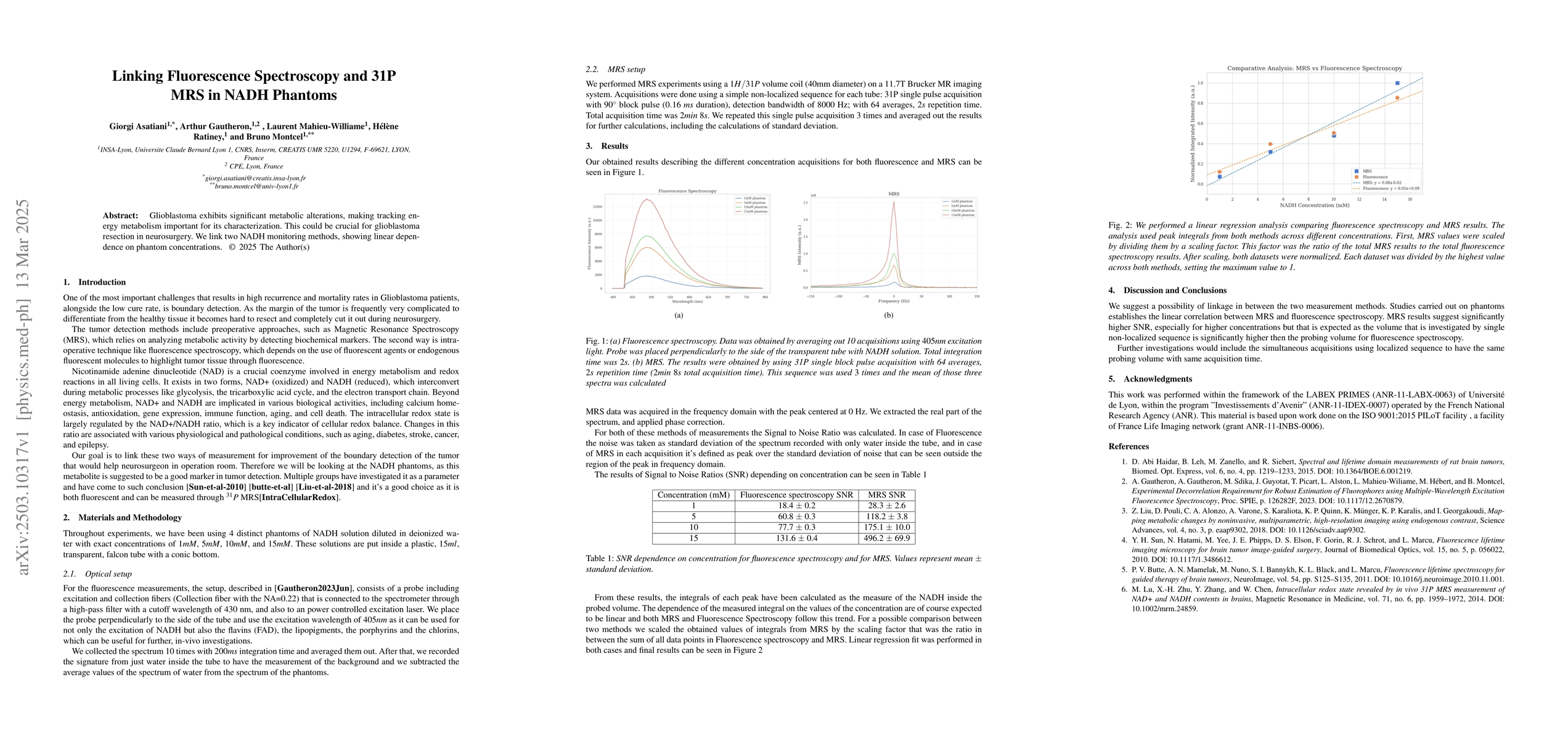
The study utilized four NADH phantoms with concentrations of 1mM, 5mM, 10mM, and 15mM in deionized water, measured using both fluorescence spectroscopy and 31P MRS techniques.
Discussion 0