Publication
Metrics
AI Quick Summary
This paper investigates the signal formation mechanisms in electrochemical strain microscopy (ESM) and provides an analytical description of its operation in frequency and time domains. It compares ESM spectroscopic modes to classical electrochemical methods, demonstrating the potential for spatially resolved studies of Li-ion dynamics at sub-10 nanometer resolution.
Paper Preview
Abstract
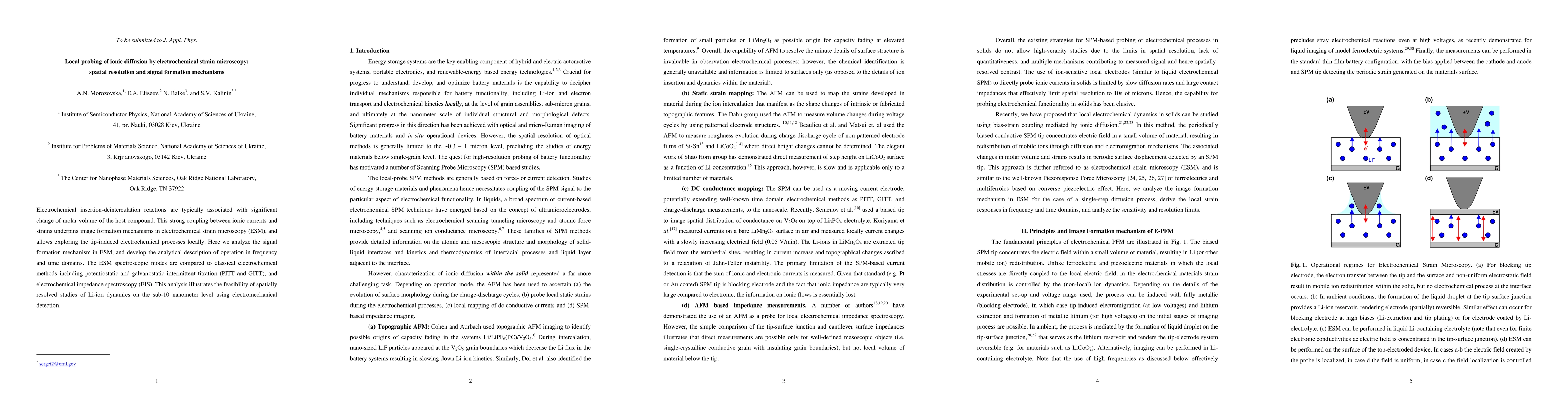
Electrochemical insertion-deintercalation reactions are typically associated with significant change of molar volume of the host compound. This strong coupling between ionic currents and strains underpins image formation mechanisms in electrochemical strain microscopy (ESM), and allows exploring the tip-induced electrochemical processes locally. Here we analyze the signal formation mechanism in ESM, and develop the analytical description of operation in frequency and time domains. The ESM spectroscopic modes are compared to classical electrochemical methods including potentiostatic and galvanostatic intermittent titration (PITT and GITT), and electrochemical impedance spectroscopy (EIS). This analysis illustrates the feasibility of spatially resolved studies of Li-ion dynamics on the sub-10 nanometer level using electromechanical detection.
AI Key Findings
Get AI-generated insights about this paper's methodology, results, significance, and more — seven facets brought into focus.
Impact
Paper Details
PDF Preview
Key Terms
Citation Network
Current paper (gray), citations (green), references (blue)
Display is limited for performance on very large graphs.
Discussion 0