Lorentz Force Electrical Impedance Tomography

Source: ArXiv

AI Quick Summary

This paper introduces Lorentz Force Electrical Impedance Tomography, a novel method for measuring the electrical conductivity of biological tissues via sonication in a magnetic field, revealing potential for detecting pathologies unseen by standard ultrasonography. Initial experiments on gelatin and beef samples show promising correlation between electrical conductivity and ultrasonic images.

Share: Share on X LinkedIn

Summary

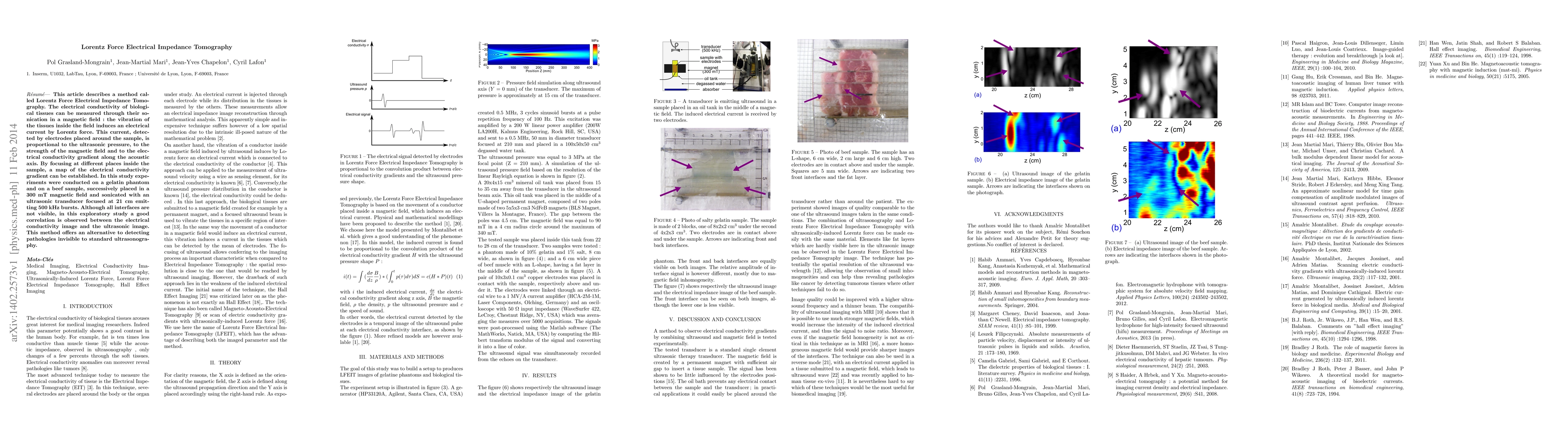
This article describes a method called Lorentz Force Electrical Impedance Tomography. The electrical conductivity of biological tissues can be measured through their sonication in a magnetic field: the vibration of the tissues inside the field induces an electrical current by Lorentz force. This current, detected by electrodes placed around the sample, is proportional to the ultrasonic pressure, to the strength of the magnetic field and to the electrical conductivity gradient along the acoustic axis. By focusing at different places inside the sample, a map of the electrical conductivity gradient can be established. In this study experiments were conducted on a gelatin phantom and on a beef sample, successively placed in a 300 mT magnetic field and sonicated with an ultrasonic transducer focused at 21 cm emitting 500 kHz bursts. Although all interfaces are not visible, in this exploratory study a good correlation is observed between the electrical conductivity image and the ultrasonic image. This method offers an alternative to detecting pathologies invisible to standard ultrasonography.

AI Key Findings

Generated Sep 07, 2025

Methodology

The research employs Lorentz Force Electrical Impedance Tomography, utilizing a magnetic field, ultrasonic transducer, and electrodes to measure the electrical conductivity of biological tissues by detecting induced electrical currents proportional to conductivity gradients and ultrasonic pressure shapes.

Key Results

  • Successful experimental testing on gelatin and beef samples in a 300 mT magnetic field and 500 kHz ultrasonic bursts.
  • Good correlation observed between the electrical conductivity image and the ultrasonic image, indicating the method's potential for pathology detection.
  • The technique offers high spatial resolution comparable to ultrasound imaging and can reveal small inhomogeneities, potentially aiding in the detection of tumors.

Significance

This research presents an alternative method for detecting pathologies invisible to standard ultrasonography, with potential applications in medical imaging and diagnostics.

Technical Contribution

The paper introduces a novel method combining ultrasonography and magnetic fields to measure electrical conductivity gradients in biological tissues, offering improved pathology detection capabilities.

Novelty

This research distinguishes itself by integrating Lorentz force principles with electrical impedance tomography and ultrasonography, providing a new approach for medical imaging and diagnostics.

Limitations

  • The current study only demonstrates the method on gelatin and beef samples; further research is needed to validate its effectiveness on human tissues.
  • Image quality could be improved with higher ultrasound frequencies and thinner beams.

Future Work

  • Explore the use of stronger magnetic fields for increased signal intensity and resolution.
  • Investigate the technique's applicability on various human tissues and organs.

Paper Details

PDF Preview

Download PDF

Key Terms

electrical (0.356)
conductivity (0.292)
placed (0.224)
lorentz (0.213)
magnetic field (0.200)
inside (0.190)
sample (0.180)
force (0.179)
Title Authors Year Actions

Comments (0)

This website uses cookies to ensure you get the best experience. Learn more