Low-Cost Continuous-Wave Diffusive Microtomography with Fiber-Scanned White-Light Illumination
Publication
Metrics
Paper Preview
Abstract
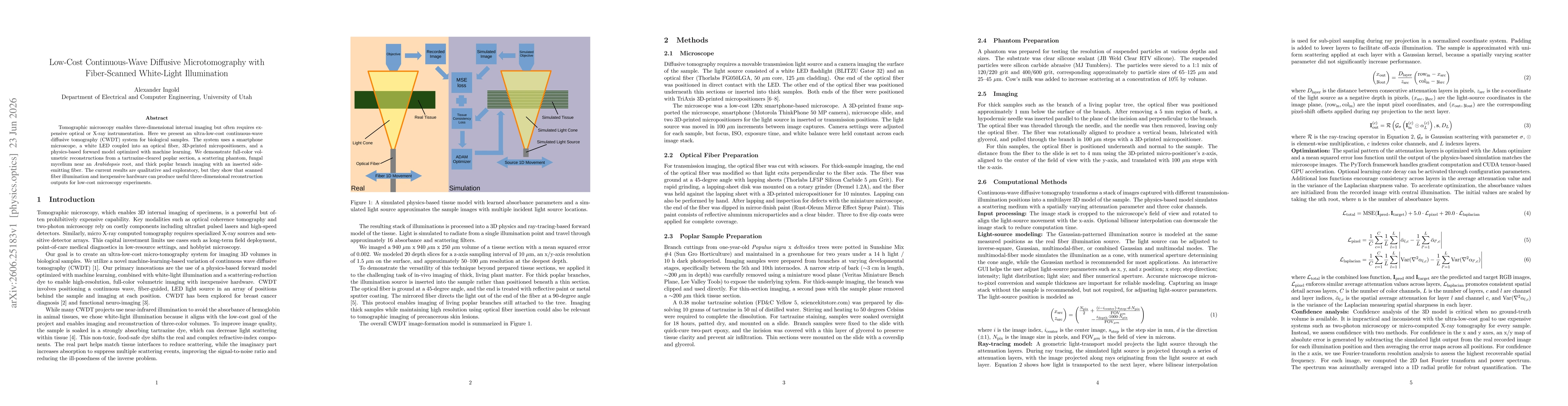
Tomographic microscopy enables three-dimensional internal imaging but often requires expensive optical or X-ray instrumentation. Here we present an ultra-low-cost continuous-wave diffusive tomography (CWDT) system for biological samples. The system uses a smartphone microscope, a white LED coupled into an optical fiber, 3D-printed micropositioners, and a physics-based forward model optimized with machine learning. We demonstrate full-color volumetric reconstructions from a tartrazine-cleared poplar section, a scattering phantom, fungal mycelium near an Arabidopsis root, and thick poplar branch imaging with an inserted side-emitting fiber. The current results are qualitative and exploratory, but they show that scanned fiber illumination and inexpensive hardware can produce useful three-dimensional reconstruction outputs for low-cost microscopy experiments.
AI Key Findings
Get AI-generated insights about this paper's methodology, results, significance, and more — seven facets brought into focus.
Discussion 0