Publication
Metrics
AI Quick Summary
This paper explores the use of the U-NET deep learning model to enhance low-dose CT images, achieving significant improvements in image quality and diagnostic preference compared to their low-dose versions, while maintaining a reduced ionizing radiation dose. Radiologists' blind tests confirm the enhanced images' superiority over both low-dose and full-dose CT images.
Paper Preview
Abstract
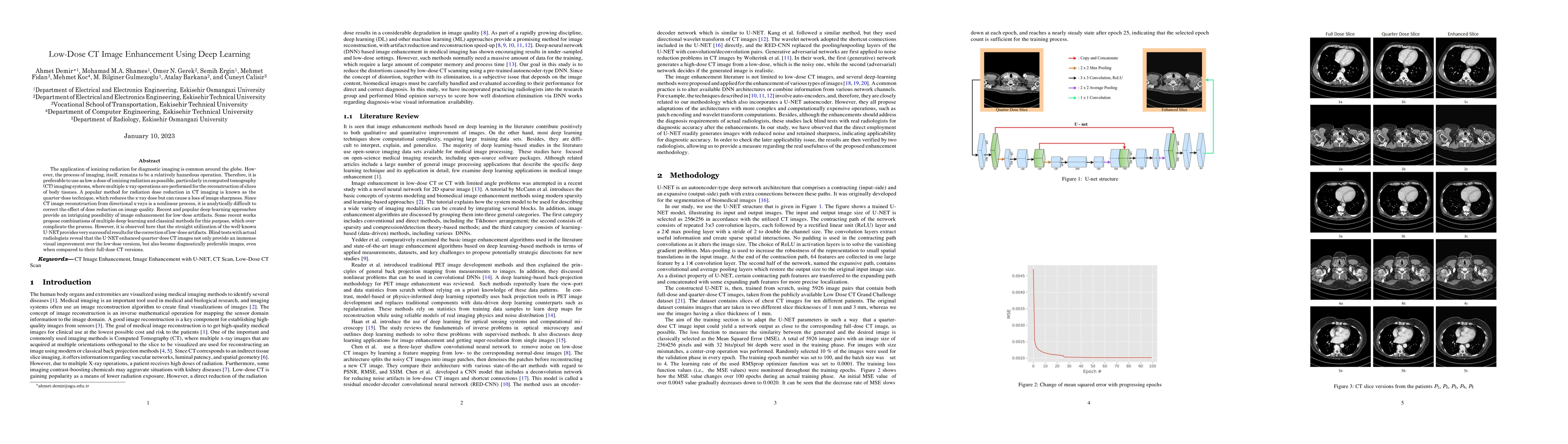
The application of ionizing radiation for diagnostic imaging is common around the globe. However, the process of imaging, itself, remains to be a relatively hazardous operation. Therefore, it is preferable to use as low a dose of ionizing radiation as possible, particularly in computed tomography (CT) imaging systems, where multiple x-ray operations are performed for the reconstruction of slices of body tissues. A popular method for radiation dose reduction in CT imaging is known as the quarter-dose technique, which reduces the x-ray dose but can cause a loss of image sharpness. Since CT image reconstruction from directional x-rays is a nonlinear process, it is analytically difficult to correct the effect of dose reduction on image quality. Recent and popular deep-learning approaches provide an intriguing possibility of image enhancement for low-dose artifacts. Some recent works propose combinations of multiple deep-learning and classical methods for this purpose, which over-complicate the process. However, it is observed here that the straight utilization of the well-known U-NET provides very successful results for the correction of low-dose artifacts. Blind tests with actual radiologists reveal that the U-NET enhanced quarter-dose CT images not only provide an immense visual improvement over the low-dose versions, but also become diagnostically preferable images, even when compared to their full-dose CT versions.
AI Key Findings
Get AI-generated insights about this paper's methodology, results, significance, and more — seven facets brought into focus.
Impact
Paper Details
Authors
PDF Preview
Key Terms
Citation Network
Current paper (gray), citations (green), references (blue)
Display is limited for performance on very large graphs.
Discussion 0