ArxivLens

Publication

Published: Aug 18, 2026
Categories: physics.med-ph

Metrics

Source: ArXiv

AI Quick Summary

LV-CARE-Diff introduces a conditional diffusion model that reconstructs the full left ventricle shape from ultra-sparse cine MRI slices (three short-axis and two long-axis) using a coarse-to-fine 3DUNet backbone plus diffusion refinement. It preserves 96% of functional accuracy while cutting scan time by 73%, enabling faster CMR exams without sacrificing quantitative measurements.

Quick Answers

What is "LV-CARE-Diff: A Conditional Anatomy-Aware Diffusion Model for Left Ventricular Shape Reconstruction and Function Quantification from Ultra-Sparse Cine Slices" about?

LV-CARE-Diff introduces a conditional diffusion model that reconstructs the full left ventricle shape from ultra-sparse cine MRI slices (three short-axis and two long-axis) using a coarse-to-fine 3DUNet backbone plus diffusion refinement. It preserves 96% of functional accuracy while cutting scan time by 73%, enabling faster CMR exams without sacrificing quantitative measurements.

What methodology did the authors use?

The study introduces LV-CARE-Diff, a coarse-to-fine pipeline that first uses a 3D U-Net to generate a coarse complete LV shape from ultra-sparse cine slices (3 SAX + 2 LAX) and then refines it with a residual diffusion model. A condition-guided input incorporating imaging plane orientation and positional metadata is used to provide spatial awareness, paired with a multi-objective training regime... More in Methodology →

What are the key results?

LV-CARE-Diff achieved the highest reconstruction performance among tested models when reconstructing complete LV shape from ultra-sparse inputs. — Deep learning models maintained about 96% of functional quantification accuracy (e.g., EDV, ESV, SV, and EF) despite sparse data, while reducing imaging time by approximately 73%. More in Key Results →

Why is this work significant?

The approach enables ultra-sparse cine acquisition, substantially shortening CMR examination time without sacrificing quantitative functional accuracy, which could enhance throughput and patient comfort in clinical workflows and large-scale screening. More in Significance →

What are the main limitations?

The study focuses on LV reconstruction from a fixed sparse set (3 SAX + 2 LAX); performance for other sparsity patterns remains untested. — Clinical validation may be needed across diverse scanner hardware and population cohorts to establish generalizability. More in Limitations →

Paper Preview

Abstract

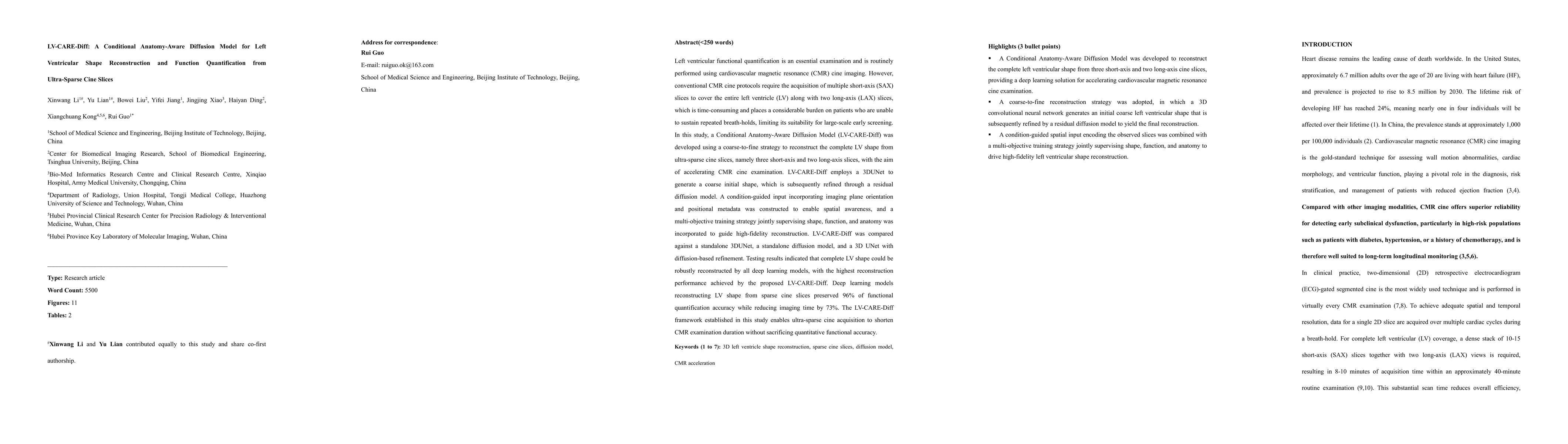
Left ventricular functional quantification is an essential examination and is routinely performed using cardiovascular magnetic resonance (CMR) cine imaging. However, conventional CMR cine protocols require the acquisition of multiple short-axis (SAX) slices to cover the entire left ventricle (LV) along with two long-axis (LAX) slices, which is time-consuming and places a considerable burden on patients who are unable to sustain repeated breath-holds, limiting its suitability for large-scale early screening. In this study, a Conditional Anatomy-Aware Diffusion Model (LV-CARE-Diff) was developed using a coarse-to-fine strategy to reconstruct the complete LV shape from ultra-sparse cine slices, namely three short-axis and two long-axis slices, with the aim of accelerating CMR cine examination. LV-CARE-Diff employs a 3DUNet to generate a coarse initial shape, which is subsequently refined through a residual diffusion model. A condition-guided input incorporating imaging plane orientation and positional metadata was constructed to enable spatial awareness, and a multi-objective training strategy jointly supervising shape, function, and anatomy was incorporated to guide high-fidelity reconstruction. LV-CARE-Diff was compared against a standalone 3DUNet, a standalone diffusion model, and a 3D UNet with diffusion-based refinement. Testing results indicated that complete LV shape could be robustly reconstructed by all deep learning models, with the highest reconstruction performance achieved by the proposed LV-CARE-Diff. Deep learning models reconstructing LV shape from sparse cine slices preserved 96% of functional quantification accuracy while reducing imaging time by 73%. The LV-CARE-Diff framework established in this study enables ultra-sparse cine acquisition to shorten CMR examination duration without sacrificing quantitative functional accuracy.
Key Findings, in focus

Seven facets of this paper, analysed and brought into focus by AI.

AI-generated · verify against source Generated Aug 20, 2026
In focus · one-line synthesis

The approach enables ultra-sparse cine acquisition, substantially shortening CMR examination time without sacrificing quantitative functional accuracy, which could enhance throughput and patient comfort in clinical workflows and large-scale screening.

01
MethodologyHow they did it

The study introduces LV-CARE-Diff, a coarse-to-fine pipeline that first uses a 3D U-Net to generate a coarse complete LV shape from ultra-sparse cine slices (3 SAX + 2 LAX) and then refines it with a residual diffusion model. A condition-guided input incorporating imaging plane orientation and positional metadata is used to provide spatial awareness, paired with a multi-objective training regime that jointly supervises shape, function, and anatomy.

02
ResultsWhat they found
  • LV-CARE-Diff achieved the highest reconstruction performance among tested models when reconstructing complete LV shape from ultra-sparse inputs.
  • Deep learning models maintained about 96% of functional quantification accuracy (e.g., EDV, ESV, SV, and EF) despite sparse data, while reducing imaging time by approximately 73%.
  • Compared with baseline methods (standalone 3D U-Net, standalone diffusion, and 3D U-Net with diffusion-based refinement), LV-CARE-Diff showed superior fidelity in both shape reconstruction and functional quantification.
03
SignificanceWhy it matters

The approach enables ultra-sparse cine acquisition, substantially shortening CMR examination time without sacrificing quantitative functional accuracy, which could enhance throughput and patient comfort in clinical workflows and large-scale screening.

04
Technical ContributionsWhat's new technically

A novel LV-CARE-Diff framework that combines a coarse 3D U-Net with a residual diffusion refinement, augmented by a condition-guided input encoding plane orientation and spatial metadata, and trained with a multi-objective loss for simultaneous shape, function, and anatomical fidelity.

05
NoveltyWhat sets it apart

Introduces a conditional anatomy-aware diffusion approach specifically designed for ultra-sparse cine slices to reconstruct complete LV morphology and quantify function, achieving high fidelity with substantially reduced imaging time.

06
LimitationsWhat to be cautious about
  • The study focuses on LV reconstruction from a fixed sparse set (3 SAX + 2 LAX); performance for other sparsity patterns remains untested.
  • Clinical validation may be needed across diverse scanner hardware and population cohorts to establish generalizability.
  • Potential sensitivity to extreme anatomical variations or pathological conditions not covered in the training data was not fully explored.
07
Future WorkWhere it goes next
  • Extend the framework to handle varying sparsity patterns and automate selection of the most informative slices.
  • Evaluate performance across larger, multi-center datasets and include more pathologies to ensure robustness.
  • Integrate user-friendly deployment tools for real-time or near-real-time clinical use and investigate integration with existing PACS workflows.

Paper Details

Paper ID: 2608.17357
Comments: 55 pages, 11 main-text figures, 2 main-text tables; supplementary material included
Categories:
physics.med-ph

How to Cite This Paper

Reference manager: RIS EndNote


Discussion 0

This website uses cookies to ensure you get the best experience. Learn more