M-Net with Bidirectional ConvLSTM for Cup and Disc Segmentation in Fundus Images
Publication
Metrics
AI Quick Summary
This paper proposes a modified M-Net with bidirectional ConvLSTM for segmenting cup and disc regions in fundus images to aid in early glaucoma detection. The model achieves a dice score of 0.92 for optic disc and 98.99% accuracy in segmenting cup and disc regions, demonstrating effective segmentation for assessing the Cup to Disc ratio.
Paper Preview
Abstract
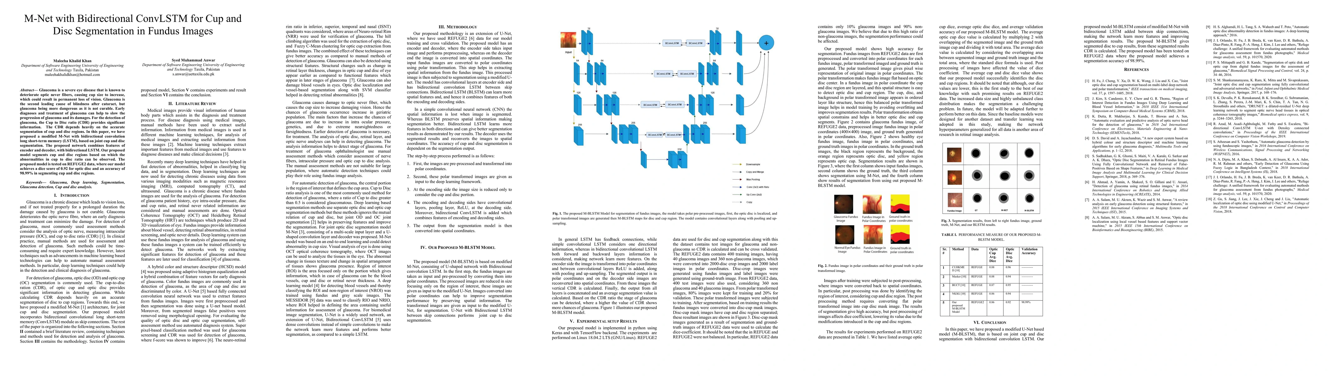
Glaucoma is a severe eye disease that is known to deteriorate optic never fibers, causing cup size to increase, which could result in permanent loss of vision. Glaucoma is the second leading cause of blindness after cataract, but glaucoma being more dangerous as it is not curable. Early diagnoses and treatment of glaucoma can help to slow the progression of glaucoma and its damages. For the detection of glaucoma, the Cup to Disc ratio (CDR) provides significant information. The CDR depends heavily on the accurate segmentation of cup and disc regions. In this paper, we have proposed a modified M-Net with bidirectional convolution long short-term memory (LSTM), based on joint cup and disc segmentation. The proposed network combines features of encoder and decoder, with bidirectional LSTM. Our proposed model segments cup and disc regions based on which the abnormalities in cup to disc ratio can be observed. The proposed model is tested on REFUGE2 data, where our model achieves a dice score of 0.92 for optic disc and an accuracy of 98.99% in segmenting cup and disc regions
AI Key Findings
Get AI-generated insights about this paper's methodology, results, significance, and more — seven facets brought into focus.
Impact
Paper Details
Authors
PDF Preview
Key Terms
Citation Network
Current paper (gray), citations (green), references (blue)
Display is limited for performance on very large graphs.
Discussion 0