Machine Friendly Machine Learning: Interpretation of Computed Tomography Without Image Reconstruction
Publication
Metrics
AI Quick Summary
This paper introduces SinoNet, a deep learning system for directly processing raw computed tomography data in sinogram-space, bypassing image reconstruction. SinoNet performed favorably for body region identification and intracranial hemorrhage detection, especially with sparse sinograms, suggesting potential for low-radiation field settings and highlighting deep learning's ability to interpret complex data beyond human capability.
Paper Preview
Abstract
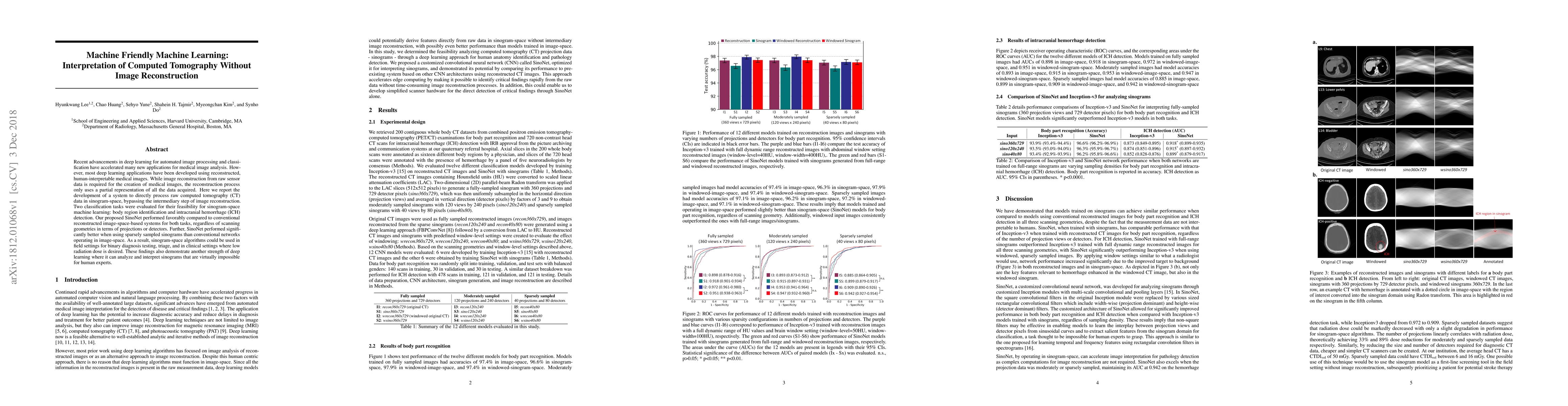
Recent advancements in deep learning for automated image processing and classification have accelerated many new applications for medical image analysis. However, most deep learning applications have been developed using reconstructed, human-interpretable medical images. While image reconstruction from raw sensor data is required for the creation of medical images, the reconstruction process only uses a partial representation of all the data acquired. Here we report the development of a system to directly process raw computed tomography (CT) data in sinogram-space, bypassing the intermediary step of image reconstruction. Two classification tasks were evaluated for their feasibility for sinogram-space machine learning: body region identification and intracranial hemorrhage (ICH) detection. Our proposed SinoNet performed favorably compared to conventional reconstructed image-space-based systems for both tasks, regardless of scanning geometries in terms of projections or detectors. Further, SinoNet performed significantly better when using sparsely sampled sinograms than conventional networks operating in image-space. As a result, sinogram-space algorithms could be used in field settings for binary diagnosis testing, triage, and in clinical settings where low radiation dose is desired. These findings also demonstrate another strength of deep learning where it can analyze and interpret sinograms that are virtually impossible for human experts.
AI Key Findings
Get AI-generated insights about this paper's methodology, results, significance, and more — seven facets brought into focus.
Impact
Paper Details
PDF Preview
Key Terms
Citation Network
Current paper (gray), citations (green), references (blue)
Display is limited for performance on very large graphs.
Discussion 0