MAD-AD: Masked Diffusion for Unsupervised Brain Anomaly Detection

Source: ArXiv

AI Quick Summary

This paper introduces a novel approach, Masked Diffusion for Unsupervised Brain Anomaly Detection (MAD-AD), which leverages masked diffusion models to learn robust representations of normal brain anatomy from MRI scans. The method identifies and localizes anomalies by detecting noisy patches in the latent space, outperforming existing techniques in anomaly detection.

Share: Share on X LinkedIn

Summary

Unsupervised anomaly detection in brain images is crucial for identifying injuries and pathologies without access to labels. However, the accurate localization of anomalies in medical images remains challenging due to the inherent complexity and variability of brain structures and the scarcity of annotated abnormal data. To address this challenge, we propose a novel approach that incorporates masking within diffusion models, leveraging their generative capabilities to learn robust representations of normal brain anatomy. During training, our model processes only normal brain MRI scans and performs a forward diffusion process in the latent space that adds noise to the features of randomly-selected patches. Following a dual objective, the model learns to identify which patches are noisy and recover their original features. This strategy ensures that the model captures intricate patterns of normal brain structures while isolating potential anomalies as noise in the latent space. At inference, the model identifies noisy patches corresponding to anomalies and generates a normal counterpart for these patches by applying a reverse diffusion process. Our method surpasses existing unsupervised anomaly detection techniques, demonstrating superior performance in generating accurate normal counterparts and localizing anomalies. The code is available at hhttps://github.com/farzad-bz/MAD-AD.
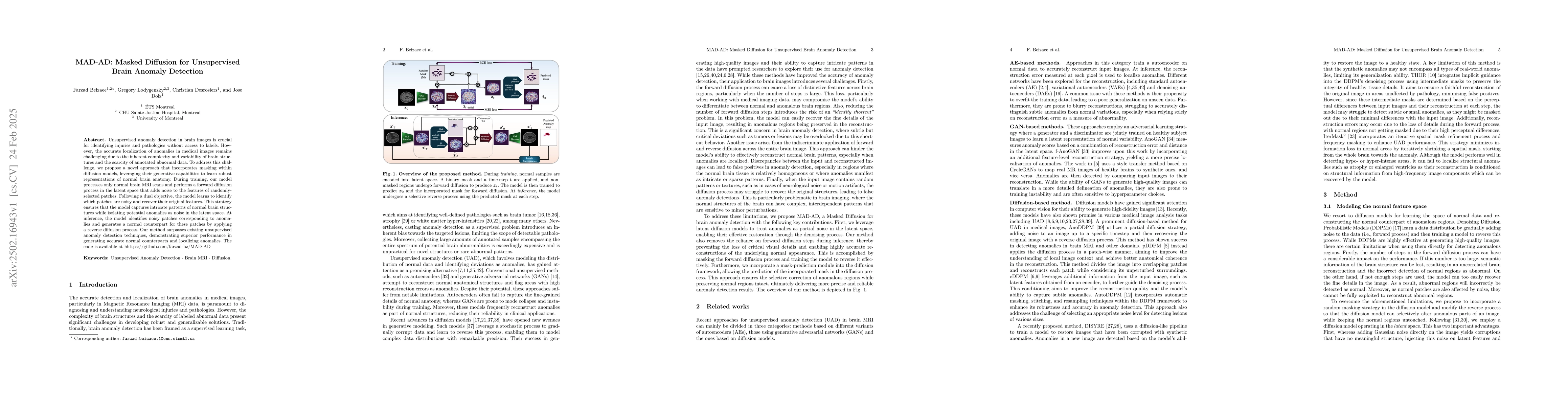
AI Key Findings

Generated Jun 11, 2025

Methodology

The research proposes MAD-AD, a novel unsupervised anomaly detection method for brain MRI using a latent diffusion model with random masking. It leverages the latent space where anomalies are considered noise, removable during denoising. A mask prediction module in the diffusion model selectively modifies anomalous regions while preserving normal areas.

Key Results

  • MAD-AD outperforms existing diffusion-based methods, especially for small and medium-sized lesions, by 4.0% and 10.9% respectively, and by 21.9% on average compared to the best baseline (THOR).
  • The method achieves the best scores in three out of four modalities under setting S2, leading to the highest average score, demonstrating its superior performance in unsupervised brain anomaly detection.
  • Ablation studies show that using image-level differences for anomaly score yields better performance than pixel-level or latent-space discrepancies.

Significance

This research is significant as it introduces an effective unsupervised anomaly detection method for brain MRI, which is crucial for identifying injuries and pathologies without labeled data. The method's ability to accurately localize anomalies without supervised labels showcases its potential as a powerful alternative to existing methods.

Technical Contribution

The key technical contribution is the use of a masked diffusion model in the latent space for unsupervised anomaly detection, combined with a mask prediction module to selectively modify anomalous regions while preserving normal areas.

Novelty

MAD-AD introduces a novel approach by incorporating random masking within diffusion models to learn robust representations of normal brain anatomy, surpassing existing unsupervised anomaly detection techniques in accuracy and anomaly localization.

Limitations

  • The model may struggle to accurately locate very small abnormalities due to the use of a diffusion model on a compressed latent space.
  • Performance evaluation is limited to specific datasets (IXI, ATLAS2.0, and BraTS’21) and may not generalize to other brain imaging modalities or pathologies without further validation.

Future Work

  • Investigate the method's performance on other brain imaging modalities and pathologies.
  • Explore techniques to improve the detection of very small anomalies in the latent space.

Paper Details

PDF Preview

Download PDF

Citation Network

Current paper (gray), citations (green), references (blue)

Display is limited for performance on very large graphs.

Current Paper
Citations
References
Click to view

No citations found for this paper.

Comments (0)

This website uses cookies to ensure you get the best experience. Learn more