Made to measure: an introduction to quantification in microscopy data
Publication
Metrics
AI Quick Summary
This paper reviews three main types of quantitative data extracted from microscopy images: intensity, morphology, and object counts, discussing their sources, measurement methods, and relevance to biological experiments. It emphasizes the importance of aligning measurements with specific biological questions to ensure valid conclusions from bioimage analysis.
Paper Preview
Abstract
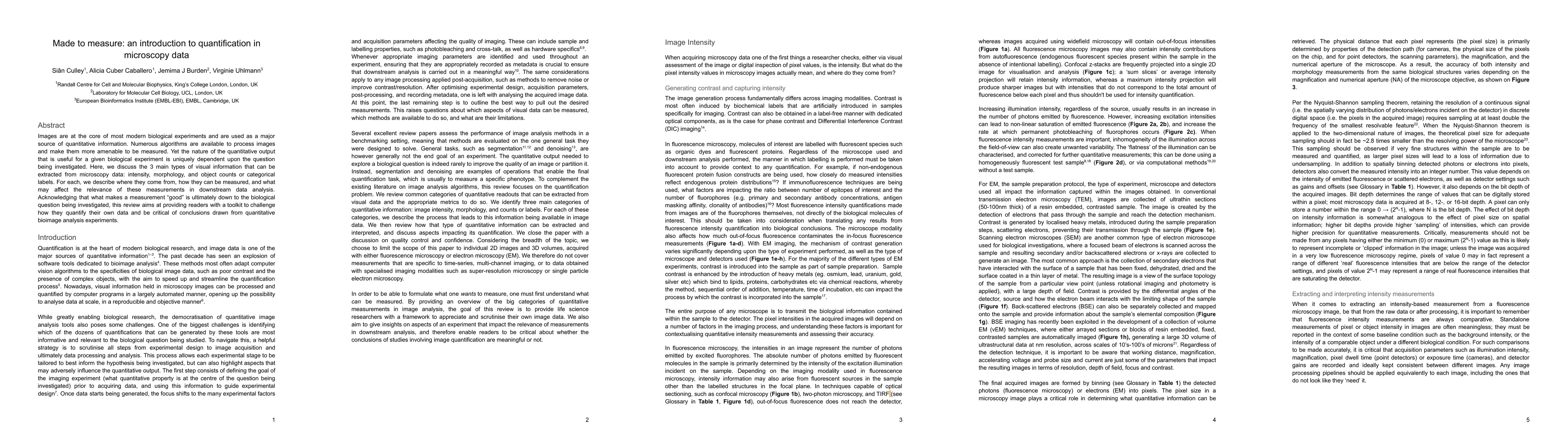
Images are at the core of most modern biological experiments and are used as a major source of quantitative information. Numerous algorithms are available to process images and make them more amenable to be measured. Yet the nature of the quantitative output that is useful for a given biological experiment is uniquely dependent upon the question being investigated. Here, we discuss the 3 main types of visual information that can be extracted from microscopy data: intensity, morphology, and object counts or categorical labels. For each, we describe where they come from, how they can be measured, and what may affect the relevance of these measurements in downstream data analysis. Acknowledging that what makes a measurement "good" is ultimately down to the biological question being investigated, this review aims at providing readers with a toolkit to challenge how they quantify their own data and be critical of conclusions drawn from quantitative bioimage analysis experiments.
AI Key Findings
Get AI-generated insights about this paper's methodology, results, significance, and more — seven facets brought into focus.
Impact
Paper Details
Authors
PDF Preview
Key Terms
Citation Network
Current paper (gray), citations (green), references (blue)
Display is limited for performance on very large graphs.
Discussion 0