Publication
Metrics
AI Quick Summary
This paper presents a method for imaging magnetic fields using NV centers in diamond, utilizing fluorescence detection to reconstruct vector magnetic field patterns with sub-micron resolution. The technique demonstrates imaging of AC magnetic fields generated by currents, with sensitivity of ~100 nT/\sqrt{Hz}, and explores potential bioimaging applications.
Paper Preview
Abstract
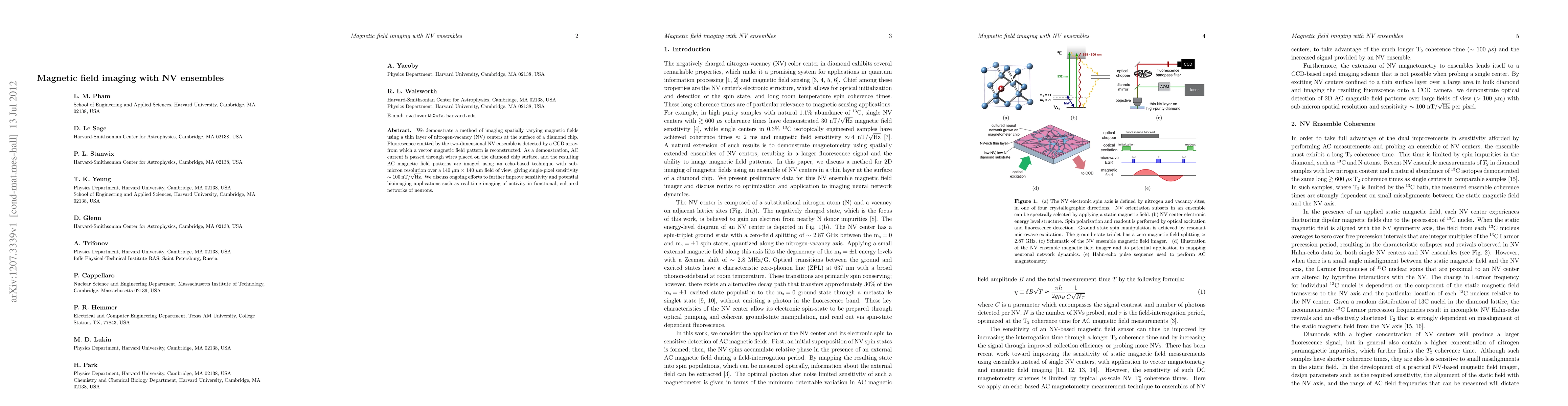
We demonstrate a method of imaging spatially varying magnetic fields using a thin layer of nitrogen-vacancy (NV) centers at the surface of a diamond chip. Fluorescence emitted by the two-dimensional NV ensemble is detected by a CCD array, from which a vector magnetic field pattern is reconstructed. As a demonstration, AC current is passed through wires placed on the diamond chip surface, and the resulting AC magnetic field patterns are imaged using an echo-based technique with sub-micron resolution over a 140 \mu m x 140 \mu m field of view, giving single-pixel sensitivity ~100 nT/\sqrt{Hz}. We discuss ongoing efforts to further improve sensitivity and potential bioimaging applications such as real-time imaging of activity in functional, cultured networks of neurons.
AI Key Findings
Get AI-generated insights about this paper's methodology, results, significance, and more — seven facets brought into focus.
Impact
Paper Details
PDF Preview
Key Terms
Citation Network
Current paper (gray), citations (green), references (blue)
Display is limited for performance on very large graphs.
Discussion 0