Purpose: Magnetization transfer (MT) and inhomogeneous MT (ihMT) contrasts
are used in MRI to provide information about macromolecular tissue content. In
particular, MT is sensitive to macromolecules and ihMT appears to be specific
to myelinated tissue. This study proposes a technique to characterize MT and
ihMT properties from a single acquisition, producing both semiquantitative
contrast ratios, and quantitative parameter maps.
Theory and Methods: Building upon previous work that uses multiband
radiofrequency (RF) pulses to efficiently generate ihMT contrast, we propose a
cyclic-steady-state approach that cycles between multiband and single-band
pulses to boost the achieved contrast. Resultant time-variable signals are
reminiscent of a magnetic resonance fingerprinting (MRF) acquisition, except
that the signal fluctuations are entirely mediated by magnetization transfer
effects. A dictionary-based low-rank inversion method is used to reconstruct
the resulting images and to produce both semiquantitative MT ratio (MTR) and
ihMT ratio (ihMTR) maps, as well as quantitative parameter estimates
corresponding to an ihMT tissue model.
Results: Phantom and in vivo brain data acquired at 1.5T demonstrate the
expected contrast trends, with ihMTR maps showing contrast more specific to
white matter (WM), as has been reported by others. Quantitative estimation of
semisolid fraction and dipolar T1 was also possible and yielded measurements
consistent with literature values in the brain.
Conclusions: By cycling between multiband and single-band pulses, an entirely
magnetization transfer mediated 'fingerprinting' method was demonstrated. This
proof-of-concept approach can be used to generate semiquantitative maps and
quantitatively estimate some macromolecular specific tissue parameters.
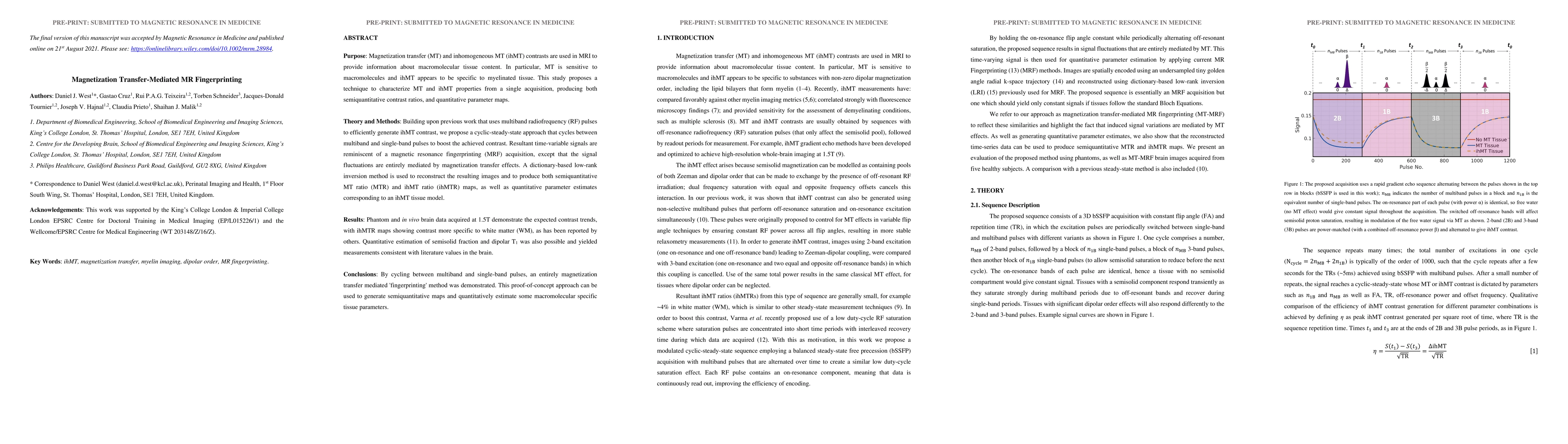
Discussion 0