Publication
Metrics
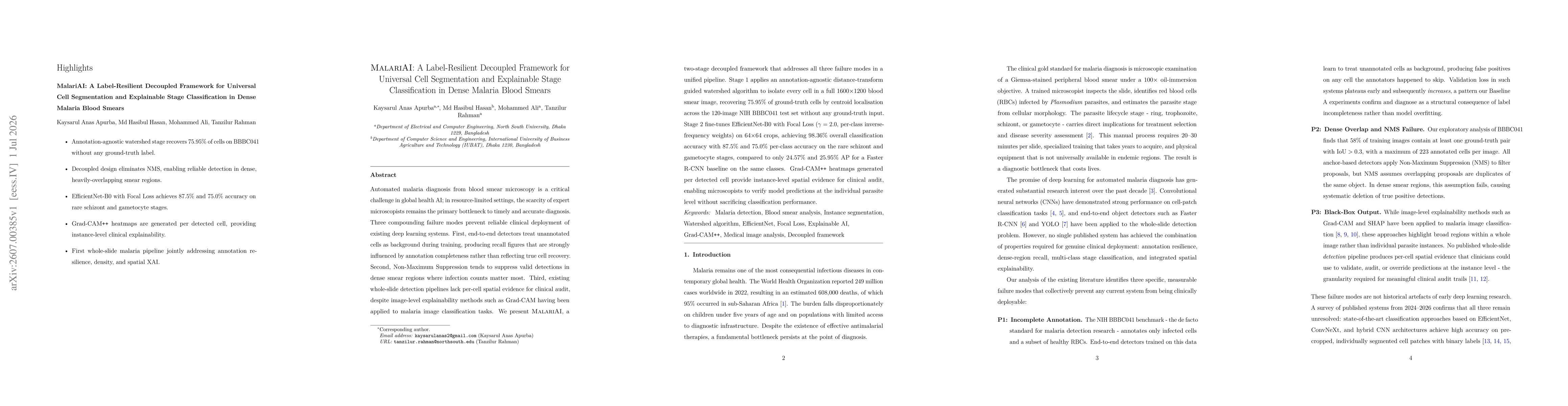
Paper Preview
Abstract
Automated malaria diagnosis from blood smear microscopy is a critical challenge in global health AI; in resource-limited settings, the scarcity of expert microscopists remains the primary bottleneck to timely and accurate diagnosis. Three compounding failure modes prevent reliable clinical deployment of existing deep learning systems. First, end-to-end detectors treat unannotated cells as background during training, producing recall figures that are strongly influenced by annotation completeness rather than reflecting true cell recovery. Second, Non-Maximum Suppression tends to suppress valid detections in dense smear regions where infection counts matter most. Third, existing whole-slide detection pipelines lack per-cell spatial evidence for clinical audit, despite image-level explainability methods such as Grad-CAM having been applied to malaria image classification tasks. We present MalariAI, a two-stage decoupled framework that addresses all three failure modes in a unified pipeline. Stage 1 applies an annotation-agnostic distance-transform guided watershed algorithm to isolate every cell in a full 1600x1200 blood smear image, recovering 75.95% of ground-truth cells by centroid localisation across the 120-image NIH BBBC041 test set without any ground-truth input. Stage 2 fine-tunes EfficientNet-B0 with Focal Loss (gamma = 2.0, per-class inverse-frequency weights) on 64x64 crops, achieving 98.36% overall classification accuracy with 87.5% and 75.0% per-class accuracy on the rare schizont and gametocyte stages, compared to only 24.57% and 25.95% AP for a Faster R-CNN baseline on the same classes. Grad-CAM++ heatmaps generated per detected cell provide instance-level spatial evidence for clinical audit, enabling microscopists to verify model predictions at the individual parasite level without sacrificing classification performance.
AI Key Findings
Get AI-generated insights about this paper's methodology, results, significance, and more — seven facets brought into focus.
Discussion 0