Mapping cellular magnesium using X-ray microfluorescence and atomic force microscopy
1007.1744
Published Jul 13, 2010
Publication
Published:
Jul 13, 2010
Updated:
Jun 01, 2025
Categories:
q-bio.CB, physics.med-ph
Metrics
Source:
ArXiv
Paper Preview
Abstract
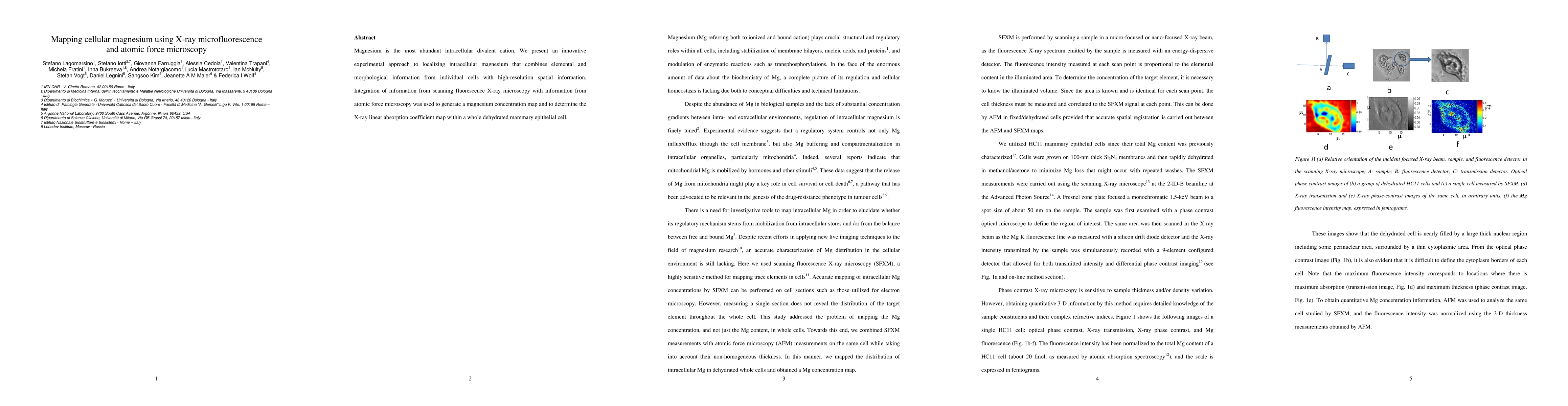
Magnesium is the most abundant intracellular divalent cation. We present an innovative experimental approach to localizing intracellular magnesium that combines elemental and morphological information from individual cells with high-resolution spatial information. Integration of information from scanning fluorescence X-ray microscopy with information from atomic force microscopy was used to generate a magnesium concentration map and to determine the X-ray linear absorption coefficient map within a whole dehydrated mammary epithelial cell.
AI Key Findings
Get AI-generated insights about this paper's methodology, results, significance, and more — seven facets brought into focus.
Paper Details
Paper ID:
1007.1744
License:
http://arxiv.org/licenses/nonexclusive-distrib/1.0/
Comments:
9 pages, 2 figures
Categories:
q-bio.CB
physics.med-ph
PDF Preview
Key Terms
microscopy
(0.333)
information
(0.278)
map
(0.270)
ray
(0.244)
fluorescence
(0.227)
morphological
(0.225)
innovative
(0.209)
scanning
(0.209)
Related Papers
No references found for this paper.
Discussion 0