Quick Summary:

This paper reviews the use of super-resolution microscopy and single-particle analysis to map supramolecular complexes, highlighting how these techniques resolve ultrastructural details at the nanoscale and provide comprehensive structural insights, overcoming limitations of underlabelling. The combination of these methods enhances resolution and coverage, broadening our understanding of biological structures.

Summary

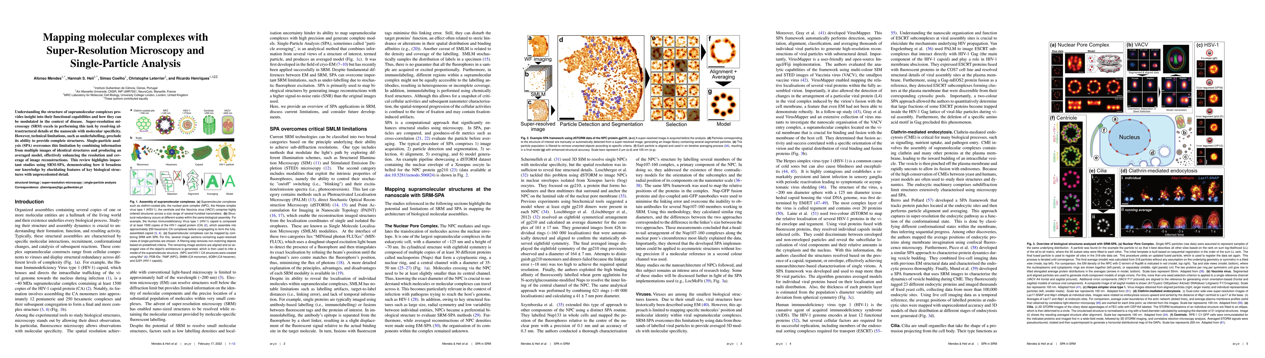
Understanding the structure of supramolecular complexes provides insight into their functional capabilities and how they can be modulated in the context of disease. Super-resolution microscopy (SRM) excels in performing this task by resolving ultrastructural details at the nanoscale with molecular specificity. However, technical limitations, such as underlabelling, preclude its ability to provide complete structures. Single-particle analysis (SPA) overcomes this limitation by combining information from multiple images of identical structures and producing an averaged model, effectively enhancing the resolution and coverage of image reconstructions. This review highlights important studies using SRM-SPA, demonstrating how it broadens our knowledge by elucidating features of key biological structures with unprecedented detail.

AI Key Findings

Get AI-generated insights about this paper's methodology, results, and significance.

Paper Details

PDF Preview

Download PDF

Key Terms

structures (0.232)
resolution (0.189)
super resolution (0.176)
unprecedented (0.172)
single particle (0.172)
nanoscale (0.171)
modulated (0.168)
limitation (0.167)
highlights (0.167)
coverage (0.166)
complexes (0.162)
producing (0.162)
disease (0.162)
biological (0.157)
averaged (0.157)
demonstrating (0.157)
identical (0.155)
microscopy (0.155)
enhancing (0.153)
technical (0.152)

Citation Network

Current paper (gray), citations (green), references (blue)

Current Paper
Citations
References
Click to view
Title Authors Year Actions
Advances in Cellular and Molecular Biology Assays: A Review of Gold Standard Methods
Katiana Simões Lopes Kittelson, Allana Cristina Faustino Martins, Gizele Celante, Arquimedes Gasparotto Júnior, Roberto Da Silva Gomes
2025
View Paper
The fundamental role of mitochondria–endoplasmic reticulum contacts in ageing and declining healthspan
Richard M. Monaghan
2025
View Paper
Structural Repetition Detector: multi-scale quantitative mapping of molecular complexes through microscopy
Afonso H. T. Mendes, Bruno M. Saraiva, Guillaume Jacquemet, J. Mamede, Christophe Leterrier, R. Henriques
2024
View Paper
Machine Learning for Single-Molecule Localization Microscopy: From Data Analysis to Quantification.
Jianli Liu, Yumian Li, Tailong Chen, Fa Zhang, Fan Xu
2024
View Paper
Single-molecule digital sizing of proteins in solution
G. Krainer, R. Jacquat, Matthias M. Schneider, Timothy J. Welsh, Jieyuan Fan, Quentin A. E. Peter, Ewa A Andrzejewska, Greta Šneiderienė, Magdalena A. Czekalska, Hannes Ausserwoeger, Lin Chai, William E. Arter, K. Saar, T. Herling, Titus M. Franzmann, V. Kosmoliaptsis, S. Alberti, F. Hartl, Steven F. Lee, T. Knowles
2024
View Paper
Decoding life's inner workings: advances in quantitative bioimaging
Ricardo Henriques, Christophe Leterrier, A. Weigel
2023
View Paper
Introducing Diinamic, a flexible and robust method for clustering analysis in single-molecule localization microscopy
Anne-Lise Paupiah, X. Marques, Zaha Merlaud, Marion Russeau, S. Lévi, M. Renner
2023
View Paper
Fast and Robust Single Particle Reconstruction in 3D Fluorescence Microscopy
Thibaut Eloy, É. Baudrier, Marine. H. Laporte, V. Hamel, P. Guichard, Denis Fortun
2023
View Paper
Reconstructing heterogeneous conformations in single-particle fluorescence imaging: application to centriole growth
View Paper

Comments (0)

This website uses cookies to ensure you get the best experience. Learn more