Mapping the spatiotemporal dynamics of calcium signaling in cellular neural networks using optical flow
Publication
Metrics
AI Quick Summary
This paper applies an optical flow gradient algorithm to map the spatiotemporal dynamics of calcium signaling in cellular neural networks with single-pixel resolution. The algorithm successfully tracks calcium signaling patterns and compares computed vector fields with manually estimated vectors, demonstrating its effectiveness across different cell types.
Paper Preview
Abstract
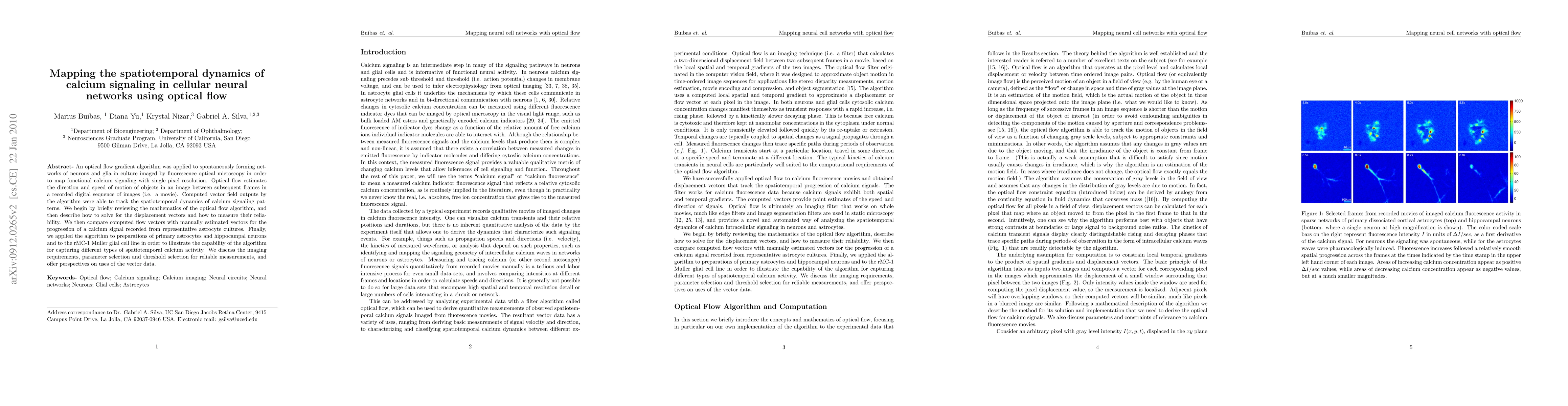
An optical flow gradient algorithm was applied to spontaneously forming net- works of neurons and glia in culture imaged by fluorescence optical microscopy in order to map functional calcium signaling with single pixel resolution. Optical flow estimates the direction and speed of motion of objects in an image between subsequent frames in a recorded digital sequence of images (i.e. a movie). Computed vector field outputs by the algorithm were able to track the spatiotemporal dynamics of calcium signaling pat- terns. We begin by briefly reviewing the mathematics of the optical flow algorithm, and then describe how to solve for the displacement vectors and how to measure their reliability. We then compare computed flow vectors with manually estimated vectors for the progression of a calcium signal recorded from representative astrocyte cultures. Finally, we applied the algorithm to preparations of primary astrocytes and hippocampal neurons and to the rMC-1 Muller glial cell line in order to illustrate the capability of the algorithm for capturing different types of spatiotemporal calcium activity. We discuss the imaging requirements, parameter selection and threshold selection for reliable measurements, and offer perspectives on uses of the vector data.
AI Key Findings
Get AI-generated insights about this paper's methodology, results, significance, and more — seven facets brought into focus.
Impact
Paper Details
PDF Preview
Key Terms
Citation Network
Current paper (gray), citations (green), references (blue)
Display is limited for performance on very large graphs.
Discussion 0