Mapping Whole Exome Sequencing to In Vivo Imaging with Stereotactic Localization and Deep Learning
Publication
Metrics
AI Quick Summary
This study combines stereotactic biopsy, MR imaging, and whole-exome sequencing to predict glioma tumor characteristics using machine learning. The approach achieved high AUC values, suggesting potential for non-invasive prediction of exome-wide mutations, which could refine targeted glioma therapies.
Paper Preview
Abstract
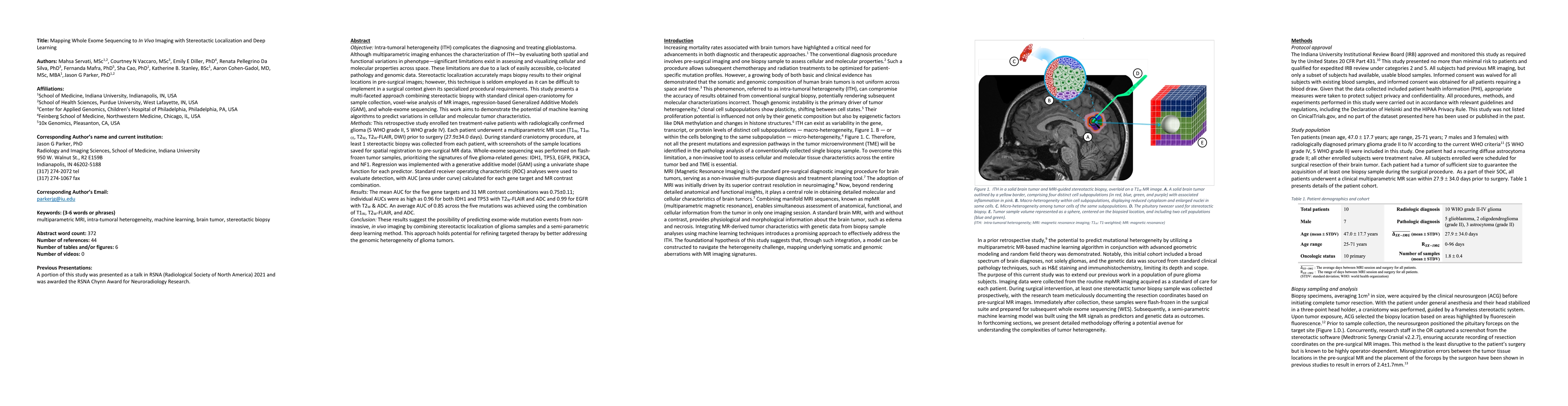
This study presents a multi-faceted approach combining stereotactic biopsy with standard clinical open-craniotomy for sample collection, voxel-wise analysis of MR images, regression-based Generalized Additive Models (GAM), & whole-exome sequencing. This work aims to demonstrate the potential of machine learning algorithms to predict variations in cellular & molecular tumor characteristics. This retrospective study enrolled ten treatment-naive patients with radiologically confirmed glioma (5 WHO grade II, 5 WHO grade IV). Each patient underwent a multiparametric MR scan (T1W, T1W-CE, T2W, T2W-FLAIR, DWI) prior to surgery (27.9+/-34.0 days). During standard craniotomy procedure, at least 1 stereotactic biopsy was collected from each patient, with screenshots of the sample locations saved for spatial registration to pre-surgical MR data. Whole-exome sequencing was performed on flash-frozen tumor samples, prioritizing the signatures of five glioma-related genes: IDH1, TP53, EGFR, PIK3CA, & NF1. Regression was implemented with a GAM using a univariate shape function for each predictor. Standard receiver operating characteristic analyses were used to evaluate detection, with AUC (area under curve) calculated for each gene target & MR contrast combination. The mean AUC for the five gene targets & 31 MR contrast combinations was 0.75+/-0.11; individual AUCs were as high as 0.96 for both IDH1 & TP53 with T2W-FLAIR & ADC & 0.99 for EGFR with T2W & ADC. An average AUC of 0.85 across the five mutations was achieved using the combination of T1W, T2W-FLAIR, & ADC. These results suggest the possibility of predicting exome-wide mutation events from non-invasive, in vivo imaging by combining stereotactic localization of glioma samples & a semi-parametric deep learning method. This approach holds potential for refining targeted therapy by better addressing the genomic heterogeneity of glioma tumors.
AI Key Findings
Get AI-generated insights about this paper's methodology, results, significance, and more — seven facets brought into focus.
Paper Details
Authors
PDF Preview
Key Terms
Related Papers
No references found for this paper.
Discussion 0