Markov Random Field Segmentation of Brain MR Images
Publication
Metrics
AI Quick Summary
This paper presents a fully-automatic 3D segmentation technique for brain MR images using Markov random fields, which effectively captures tissue intensity distributions, neighborhood correlations, and signal inhomogeneities. The method is validated through simulations and real MR images, demonstrating robust classification of different tissue types despite noise and inhomogeneities.
Paper Preview
Abstract
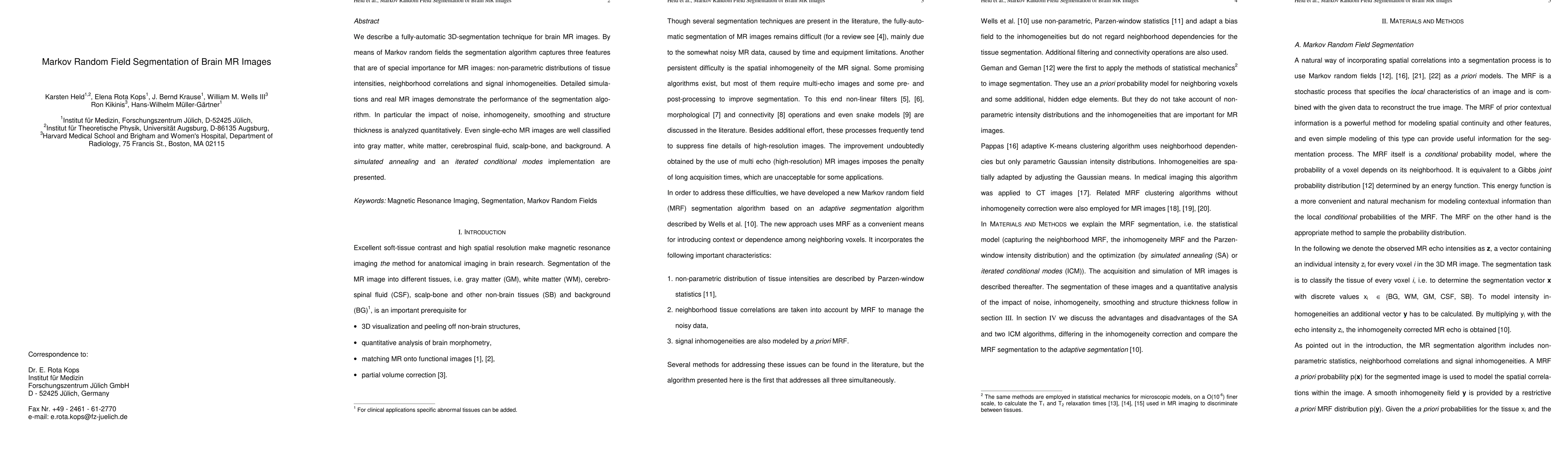
We describe a fully-automatic 3D-segmentation technique for brain MR images. Using Markov random fields the segmentation algorithm captures three important MR features, i.e. non-parametric distributions of tissue intensities, neighborhood correlations and signal inhomogeneities. Detailed simulations and real MR images demonstrate the performance of the segmentation algorithm. The impact of noise, inhomogeneity, smoothing and structure thickness is analyzed quantitatively. Even single echo MR images are well classified into gray matter, white matter, cerebrospinal fluid, scalp-bone and background. A simulated annealing and an iterated conditional modes implementation are presented. Keywords: Magnetic Resonance Imaging, Segmentation, Markov Random Fields
AI Key Findings
Get AI-generated insights about this paper's methodology, results, significance, and more — seven facets brought into focus.
Impact
Paper Details
PDF Preview
Key Terms
Citation Network
Current paper (gray), citations (green), references (blue)
Display is limited for performance on very large graphs.
Discussion 0