ArxivLens

Publication

Published: Feb 24, 2026
Categories: cs.CV

Metrics

Source: ArXiv

Quick Answers

What methodology did the authors use?

Mask-HybridGNet trains graph-based segmentation models directly from pixel-wise masks using Chamfer distance supervision, edge-based regularization, and differentiable rasterization, while enforcing a fixed adjacency matrix to maintain topological consistency More in Methodology →

What are the key results?

Achieves competitive Dice, Hausdorff, and ASSD scores versus nnUNet across chest X-ray, cardiac ultrasound, cardiac MRI, and fetal imaging datasets — Automatically learns consistent anatomical landmark correspondences enabling temporal tracking, cross-slice reconstruction, and population shape analysis without explicit landmark supervision More in Key Results →

Why is this work significant?

Provides a practical, annotation-free method to obtain graph-based, topologically sound segmentations with implicit anatomical correspondences, facilitating clinical workflows, longitudinal studies, and shape-based research More in Significance →

What are the main limitations?

Limited to 2D image segmentation; extension to 3D volumetric data remains an open problem — Requires simple closed-boundary topology; cannot directly model branching structures or holes without further graph representation extensions More in Limitations →

Paper Preview

Abstract

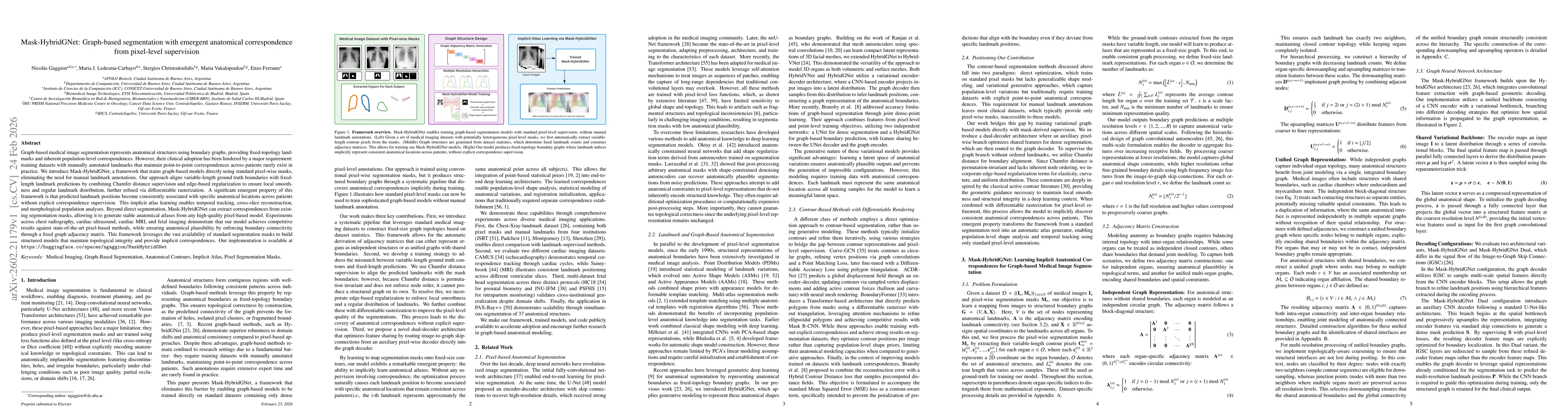
Graph-based medical image segmentation represents anatomical structures using boundary graphs, providing fixed-topology landmarks and inherent population-level correspondences. However, their clinical adoption has been hindered by a major requirement: training datasets with manually annotated landmarks that maintain point-to-point correspondences across patients rarely exist in practice. We introduce Mask-HybridGNet, a framework that trains graph-based models directly using standard pixel-wise masks, eliminating the need for manual landmark annotations. Our approach aligns variable-length ground truth boundaries with fixed-length landmark predictions by combining Chamfer distance supervision and edge-based regularization to ensure local smoothness and regular landmark distribution, further refined via differentiable rasterization. A significant emergent property of this framework is that predicted landmark positions become consistently associated with specific anatomical locations across patients without explicit correspondence supervision. This implicit atlas learning enables temporal tracking, cross-slice reconstruction, and morphological population analyses. Beyond direct segmentation, Mask-HybridGNet can extract correspondences from existing segmentation masks, allowing it to generate stable anatomical atlases from any high-quality pixel-based model. Experiments across chest radiography, cardiac ultrasound, cardiac MRI, and fetal imaging demonstrate that our model achieves competitive results against state-of-the-art pixel-based methods, while ensuring anatomical plausibility by enforcing boundary connectivity through a fixed graph adjacency matrix. This framework leverages the vast availability of standard segmentation masks to build structured models that maintain topological integrity and provide implicit correspondences.

Key Findings, in focus

Seven facets of this paper, analysed and brought into focus by AI.

AI-generated · verify against source Generated Feb 25, 2026
In focus · one-line synthesis

Provides a practical, annotation-free method to obtain graph-based, topologically sound segmentations with implicit anatomical correspondences, facilitating clinical workflows, longitudinal studies, and shape-based research

01
MethodologyHow they did it

Mask-HybridGNet trains graph-based segmentation models directly from pixel-wise masks using Chamfer distance supervision, edge-based regularization, and differentiable rasterization, while enforcing a fixed adjacency matrix to maintain topological consistency

02
ResultsWhat they found
  • Achieves competitive Dice, Hausdorff, and ASSD scores versus nnUNet across chest X-ray, cardiac ultrasound, cardiac MRI, and fetal imaging datasets
  • Automatically learns consistent anatomical landmark correspondences enabling temporal tracking, cross-slice reconstruction, and population shape analysis without explicit landmark supervision
  • Demonstrates robust cross-dataset generalization on multi-center fetal head and pubic symphysis data, outperforming nnUNet in heterogeneous annotation settings
03
SignificanceWhy it matters

Provides a practical, annotation-free method to obtain graph-based, topologically sound segmentations with implicit anatomical correspondences, facilitating clinical workflows, longitudinal studies, and shape-based research

04
Technical ContributionsWhat's new technically

Introduces a mask-supervised graph-based segmentation framework that learns fixed-node correspondences via Chamfer distance and edge regularization, enabling implicit atlas learning without manual landmark annotations

05
NoveltyWhat sets it apart

First to train graph-based segmentation models directly from standard pixel masks, eliminating the need for manual landmark annotations while achieving emergent anatomical correspondences across diverse medical imaging modalities

06
LimitationsWhat to be cautious about
  • Limited to 2D image segmentation; extension to 3D volumetric data remains an open problem
  • Requires simple closed-boundary topology; cannot directly model branching structures or holes without further graph representation extensions
07
Future WorkWhere it goes next
  • Extend the framework to 3D volumetric segmentation and surface mesh reconstruction using Chamfer-based training on variable-length 3D contours
  • Incorporate self-supervised losses such as cycle consistency and prior anatomical landmarks to further improve correspondence quality

Paper Details

Paper ID: 2602.21179
Categories:
cs.CV

How to Cite This Paper

Reference manager: RIS EndNote


Discussion 0

This website uses cookies to ensure you get the best experience. Learn more