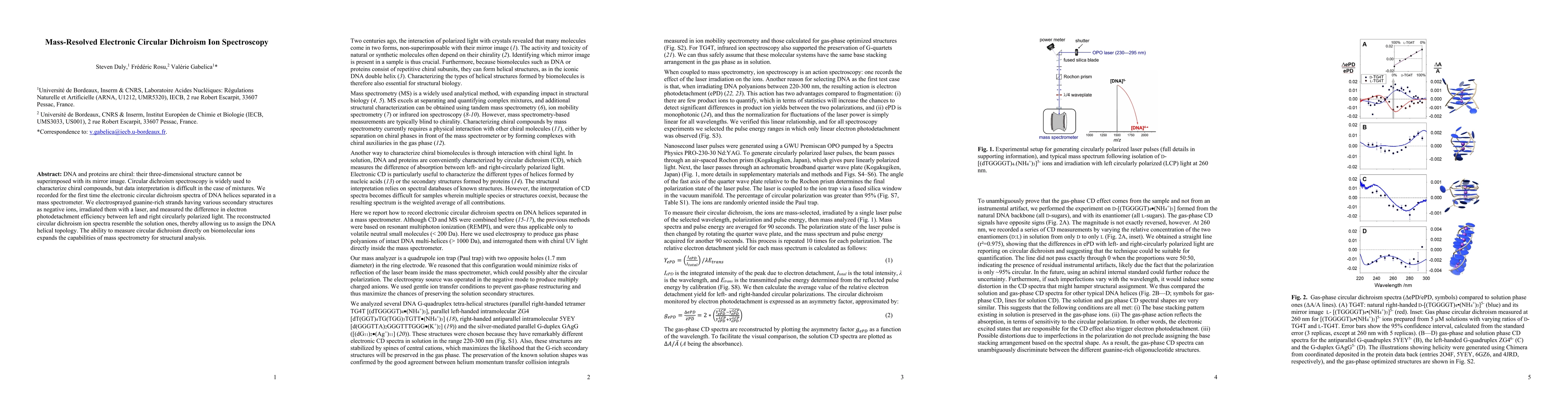
This study presents the first mass-resolved electronic circular dichroism spectroscopy of DNA helices in a mass spectrometer, enabling the characterization of chiral biomolecular ions. The technique allows for the assignment of DNA helical topology, enhancing the structural analysis capabilities of mass spectrometry.
DNA and proteins are chiral: their three-dimensional structure cannot be superimposed with its mirror image. Circular dichroism spectroscopy is widely used to characterize chiral compounds, but data interpretation is difficult in the case of mixtures. We recorded for the first time the electronic circular dichroism spectra of DNA helices separated in a mass spectrometer. We electrosprayed guanine-rich strands having various secondary structures as negative ions, irradiated them with a laser, and measured the difference in electron photodetachment efficiency between left and right circularly polarized light. The reconstructed circular dichroism ion spectra resemble the solution ones, thereby allowing us to assign the DNA helical topology. The ability to measure circular dichroism directly on biomolecular ions expands the capabilities of mass spectrometry for structural analysis.
Generated Sep 07, 2025
The research utilized mass spectrometry combined with electronic circular dichroism (CD) spectroscopy to analyze guanine-rich DNA strands with various secondary structures. Ions were produced via electrospray ionization, trapped, and irradiated with a single laser pulse. The relative electron detachment efficiency was monitored to determine CD values, which were then compared to solution-phase measurements.
This research is significant as it provides a novel approach for structural analysis of chiral biomolecules like DNA, offering a more detailed understanding of their secondary structures in the gas phase, which could aid in studying biological processes and designing therapeutic agents.
The development of mass-resolved electronic CD ion spectroscopy, which combines mass spectrometry with CD measurements to analyze chiral biomolecules in the gas phase.
This work introduces a new method for structural analysis of chiral biomolecules in the gas phase, providing CD spectra that resemble solution-phase measurements, which was not previously possible.
Current paper (gray), citations (green), references (blue)
Display is limited for performance on very large graphs.
Thomas P. Devereaux, Samuel Beaulieu, Shuo Dong et al.
Yulia Samoshkina, Dmitriy Petrov, Andrei Telegin et al.
Title | Authors | Year | Actions |
---|
Comments (0)