ArxivLens

Quick Summary:

This study presents a new method for material decomposition in x-ray imaging using phase contrast to enhance sensitivity to weakly-attenuating materials and separate overlaid materials. The single-grid x-ray phase contrast imaging technique captures data with a novel correction to remove edge-based phase effects.

DOI: 10.1364/OL.389770
arXiv Id: 2002.04417
Date Published: 2020-08-26
Date Updated: 2020-08-26

Key Terms (TF-IDF):

exposure (0.286)
material (0.216)
imaging (0.212)
contrast (0.197)
technique (0.191)
single (0.188)
ray (0.187)
phase (0.185)
high speed (0.171)
captured (0.157)
remove (0.157)
new approach (0.156)
biological (0.153)
act (0.149)
grid (0.148)
separated (0.147)
complementary (0.143)
correction (0.141)
sharp (0.141)
collected (0.138)
License: http://arxiv.org/licenses/nonexclusive-distrib/1.0/

Summary:

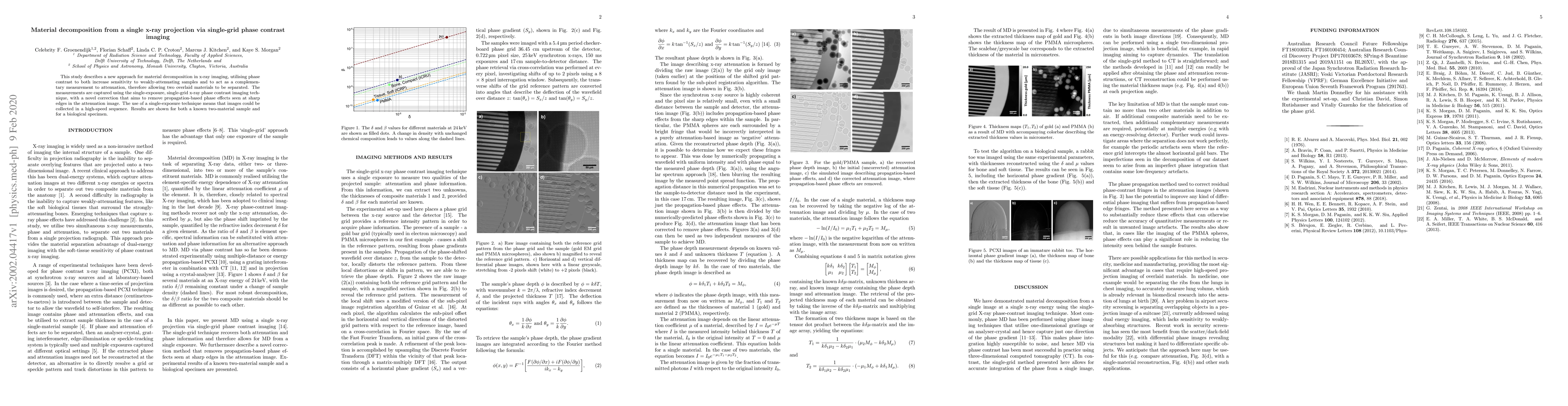
This study describes a new approach for material decomposition in x-ray imaging, utilising phase contrast to both increase sensitivity to weakly-attenuating samples and to act as a complementary measurement to attenuation, therefore allowing two overlaid materials to be separated. The measurements are captured using the single-exposure, single-grid x-ray phase contrast imaging technique, with a novel correction that aims to remove propagation-based phase effects seen at sharp edges in the attenuation image. The use of a single-exposure technique means that images could be collected in a high-speed sequence. Results are shown for both a known two-material sample and for a biological specimen.

Citation & Reference Network Visualization

This graph shows how this paper is connected to other research. The current paper is shown in dark gray, papers that cite it are in green, and papers it references are in blue. You can:

Current Paper
Papers citing this work (Citations)
Papers cited by this work (References)

• Hover over any node to see the paper title

• Click a node to view that paper's details

• Drag nodes to rearrange the graph

• Use the zoom controls (bottom right) to zoom in/out

Citations and References

Title Authors Year Actions
On the quantification of sample microstructure using single-exposure x-ray dark-field imaging via a single-grid setup
Y. Y. How, D. Paganin, K. Morgan
2022
View Paper
Directional dark-field retrieval with single-grid x-ray imaging.
M. Croughan, Y. Y. How, Allan Pennings, K. Morgan
2022
View Paper
One-Step Method for Material Quantitation Using In-Line Tomography With Single Scanning
Suyu Liao, Shiwo Deng, Yining Zhu, Huitao Zhang, P. Zhu, Kai Zhang, Xinggang Zhao
2022
View Paper
Quantifying the x-ray dark-field signal in single-grid imaging.
Y. Y. How, K. Morgan
2021
View Paper
X-Ray Dark-Field and Phase Retrieval Without Optics, via the Fokker–Planck Equation
Thomas A. Leatham, D. Paganin, K. Morgan
2021
View Paper
Directional dark-field implicit x-ray speckle tracking using an anisotropic-diffusion Fokker-Planck equation
K. Pavlov, D. Paganin, K. Morgan, H. Li, S. Berujon, Laurene Qu'enot, E. Brun
2021
View Paper
X-ray multi-modal intrinsic-speckle-tracking
K. Pavlov, D. Paganin, Heyang (Thomas) Li, S. Berujon, H'elene Roug'e-Labriet, E. Brun
2019
View Paper
Single-Shot X-Ray Speckle-Based Imaging of a Single-Material Object
K. Pavlov, H. Li, D. Paganin, S. Berujon, H'elene Roug'e-Labriet, E. Brun
2019
View Paper

Comments (0)

This website uses cookies to ensure you get the best experience. Learn more