MBIR Training for a 2.5D DL network in X-ray CT

Source: ArXiv

AI Quick Summary

This paper trains a 2.5D deep learning network, based on a modified Unet, to emulate the high-quality images of model-based iterative reconstruction (MBIR) in X-ray CT, achieving similar results faster and with lower computational costs compared to traditional MBIR methods. The DL-MBIR images show texture and reduced noise comparable to MBIR, as evidenced by NPS plots and image profile analyses.

Share: Share on X LinkedIn

Summary

In computed tomographic imaging, model based iterative reconstruction methods have generally shown better image quality than the more traditional, faster filtered backprojection technique. The cost we have to pay is that MBIR is computationally expensive. In this work we train a 2.5D deep learning (DL) network to mimic MBIR quality image. The network is realized by a modified Unet, and trained using clinical FBP and MBIR image pairs. We achieve the quality of MBIR images faster and with a much smaller computation cost. Visually and in terms of noise power spectrum (NPS), DL-MBIR images have texture similar to that of MBIR, with reduced noise power. Image profile plots, NPS plots, standard deviation, etc. suggest that the DL-MBIR images result from a successful emulation of an MBIR operator.

AI Key Findings

Generated Sep 03, 2025

Methodology

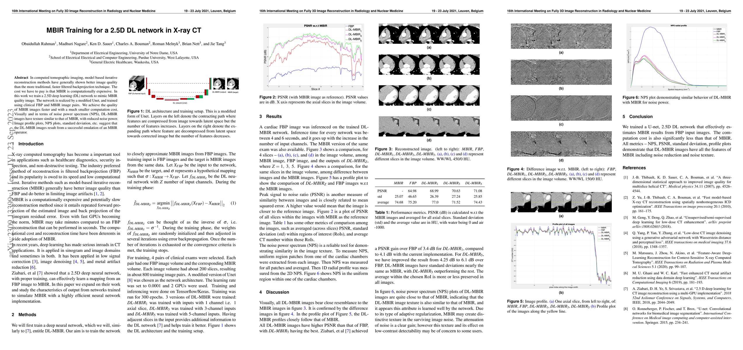
A 2.5D deep learning network, based on a modified Unet, was trained to approximate Model-Based Iterative Reconstruction (MBIR) quality images from Filtered Back Projection (FBP) images using clinical exam pairs. The network was trained with 4 pairs of clinical exams, each having one FBP image volume and the corresponding MBIR volume, resulting in about 800 training image pairs.

Key Results

  • The DL-MBIR network successfully emulated MBIR operators, producing images visually and in terms of noise power spectrum (NPS) similar to MBIR, but with reduced noise power.
  • Inferencing on the trained DL-MBIR network took between 4 to 6 seconds per cardiac slice, significantly faster than MBIR reconstruction which can take minutes.
  • DL-MBIR images had higher PSNR than FBP images, with DL-MBIR5 having the best performance, and standard deviations near MBIR, indicating similar noise reduction and texture.

Significance

This research is significant as it presents a computationally efficient alternative to MBIR for achieving high-quality X-ray CT images, which could potentially accelerate clinical workflows without compromising image quality.

Technical Contribution

The main technical contribution is the development and validation of a 2.5D deep learning network (DL-MBIR) that efficiently mimics MBIR quality images from FBP inputs, reducing computational costs significantly.

Novelty

This work expands on previous research by Ziabari et al. [7] by implementing a highly efficient neural network for simulating MBIR, offering faster reconstruction times and comparable image quality.

Limitations

  • The study was limited to cardiac imaging and did not explore its applicability to other anatomical regions.
  • The performance of the DL-MBIR network might vary with different types of clinical data and scanner configurations.

Future Work

  • Investigate the generalizability of the DL-MBIR network across various anatomical regions and scanner types.
  • Explore the integration of the DL-MBIR network into clinical practice to assess its impact on diagnostic accuracy and workflow efficiency.

Paper Details

PDF Preview

Download PDF

Key Terms

dl (0.356)
image (0.262)
quality (0.248)
image (0.246)
images (0.239)
faster (0.227)
quality (0.226)
faster (0.224)

Citation Network

Current paper (gray), citations (green), references (blue)

Display is limited for performance on very large graphs.

Current Paper
Citations
References
Click to view
Title Authors Year Actions

Comments (0)

This website uses cookies to ensure you get the best experience. Learn more