Summary
We aim to investigate the feasibility of online monitoring of irradiation time (IRT) and scan time for FLASH radiotherapy using a pixelated semiconductor detector. Measurements of the time structure of FLASH irradiations were performed using fast, pixelated spectral detectors, AdvaPIX-TPX3 and Minipix-TPX3. The latter has a fraction of its sensor coated with a neutron sensitive material. With little or no dead time and an ability to resolve events that are closely spaced in time (tens of ns), both detectors can accurately determine IRTs as long as pile-ups are avoided. To avoid pile-ups, we placed the detectors beyond the Bragg peak or at a large scattering angle. We acquired prompt gamma rays and secondary neutrons and calculated IRTs based on timestamps of the first (beam-on) and the last (beam-off) charged species. We also measured scan times in x, y, and diagonal directions. We performed these measurements for a single spot, a small animal field, a patient field, and a ridge filter optimized field to demonstrate in vivo online monitoring of IRT. All measurements were compared to vendor log files. Differences between measurements and log files for a single spot, a small animal field, and a patient field were within 1%, 0.3% and 1%, respectively. In vivo monitoring of IRTs was accurate within 0.1% for AdvaPIX-TPX3 and within 6.1% for Minipix-TPX3. The scan times in x, y, and diagonal directions were 4.0, 3.4, and 4.0 ms, respectively. Overall, the AdvaPIX-TPX3 can measure FLASH IRTs within 1% accuracy, indicating that prompt gamma rays are a good surrogate for primary protons. The Minipix-TPX3 showed a higher discrepancy, suggesting a need for further investigation. The scan times (3.4 \pm 0.05 ms) in the 60-mm distance of y-direction were less than (4.0 \pm 0.06 ms) in the 24-mm distance of x-direction, confirming the much faster scanning speed of the Y magnets than that of X.
AI Key Findings
Generated Sep 03, 2025
Methodology
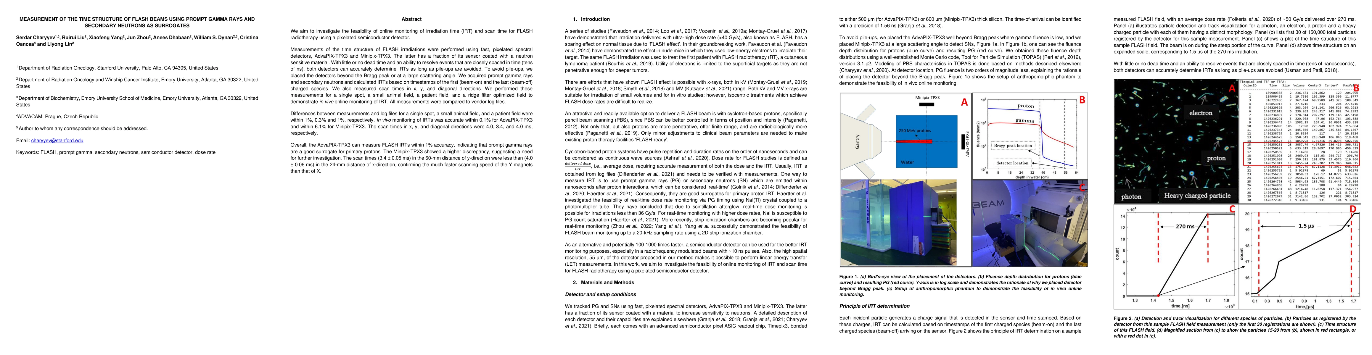
The research utilized pixelated semiconductor detectors, AdvaPIX-TPX3 and Minipix-TPX3, to measure the time structure of FLASH irradiations, avoiding pile-ups by placing detectors beyond the Bragg peak or at large scattering angles. Prompt gamma rays and secondary neutrons were acquired for IRT calculation, with timestamps used for beam-on and beam-off detection.
Key Results
- AdvaPIX-TPX3 accurately measured FLASH IRTs within 1% accuracy, indicating prompt gamma rays as a good surrogate for primary protons.
- Minipix-TPX3 showed a higher discrepancy, suggesting a need for further investigation when using secondary neutrons as surrogates.
- Scan times were measured as 4.0 ms (x-direction), 3.4 ms (y-direction), and 4.0 ms (diagonal direction), confirming faster Y magnet scanning speed than X magnets.
Significance
This study demonstrates the feasibility of online monitoring of irradiation time and scan time for FLASH radiotherapy, which is crucial for optimizing treatment delivery and ensuring accurate dose administration.
Technical Contribution
The research presents a novel approach to measure the time structure of FLASH beams using pixelated semiconductor detectors, providing a method for online monitoring of irradiation time and scan time in FLASH radiotherapy.
Novelty
The study introduces a new method for online monitoring of FLASH irradiation time and scan time using pixelated detectors, offering improved accuracy and potential for real-time dose optimization in FLASH radiotherapy.
Limitations
- Minipix-TPX3 measurements had a higher discrepancy compared to AdvaPIX-TPX3, indicating potential issues when using secondary neutrons as surrogates.
- The study did not extensively investigate the impact of various beam currents and energies on detector performance.
Future Work
- Further investigation into the performance of Minipix-TPX3 with secondary neutrons as surrogates.
- Expanding the study to include a broader range of beam currents and energies to assess detector performance and accuracy.
Paper Details
PDF Preview
Key Terms
Citation Network
Current paper (gray), citations (green), references (blue)
Display is limited for performance on very large graphs.
| Title | Authors | Year | Actions |
|---|
Comments (0)