Publication
Metrics
AI Quick Summary
This paper presents a novel method using Integrated Digital Image Correlation with Atomic Force Microscopy to measure nanoscale stress intensity factors during crack propagation in glass. The technique allows for precise quantification of stress fields down to 10 nm from the crack tip, providing accurate out-of-plane displacement measurements.
Paper Preview
Abstract
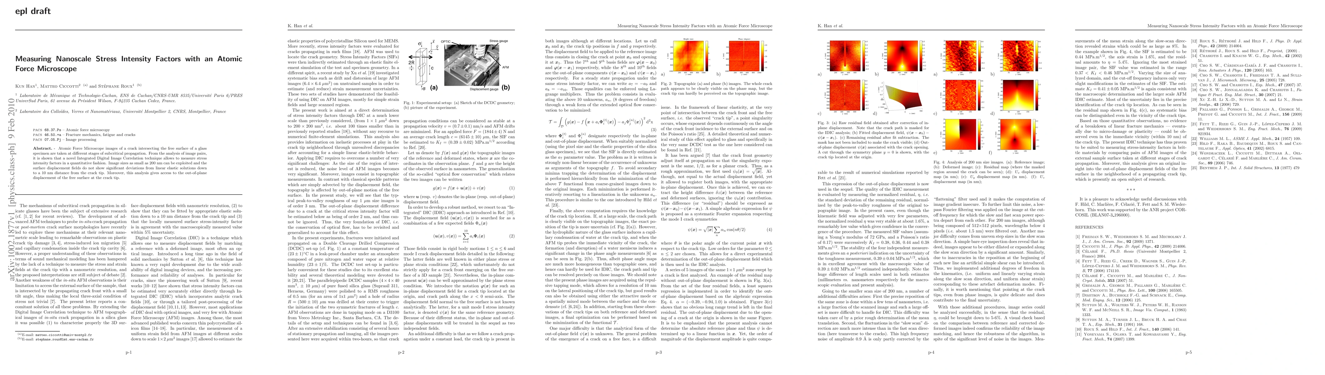
Atomic Force Microscope images of a crack intersecting the free surface of a glass specimen are taken at different stages of subcritical propagation. From the analysis of image pairs, it is shown that a novel Integrated Digital Image Correlation technique allows to measure stress intensity factors in a quantitative fashion. Image sizes as small as 200 nm can be exploited and the surface displacement fields do not show significant deviations from linear elastic solutions down to a 10 nm distance from the crack tip. Moreover, this analysis gives access to the out-of-plane displacement of the free surface at the crack tip.
AI Key Findings
Get AI-generated insights about this paper's methodology, results, significance, and more — seven facets brought into focus.
Impact
Paper Details
PDF Preview
Key Terms
Citation Network
Current paper (gray), citations (green), references (blue)
Display is limited for performance on very large graphs.
Discussion 0