Publication
Metrics
AI Quick Summary
Researchers propose a novel mechanism for T cell pattern formation during adhesion to antigen-presenting cells, involving initial microdomain nucleation and diffusion of receptors and ligands.
Paper Preview
Abstract
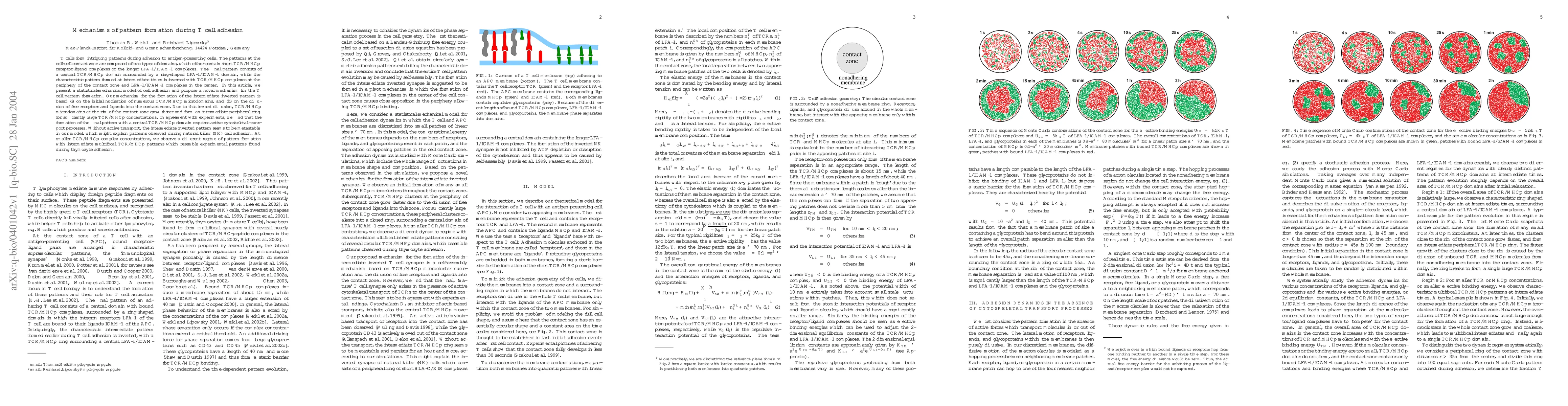
T cells form intriguing patterns during adhesion to antigen-presenting cells. The patterns at the cell-cell contact zone are composed of two types of domains, which either contain short TCR/MHCp receptor-ligand complexes or the longer LFA-1/ICAM-1 complexes. The final pattern consists of a central TCR/MHCp domain surrounded by a ring-shaped LFA-1/ICAM-1 domain, while the characteristic pattern formed at intermediate times is inverted with TCR/MHCp complexes at the periphery of the contact zone and LFA-1/ICAM-1 complexes in the center. In this article, we present a statistical-mechanical model of cell adhesion and propose a novel mechanism for the T cell pattern formation. Our mechanism for the formation of the intermediate inverted pattern is based (i) on the initial nucleation of numerous TCR/MHCp microdomains, and (ii) on the diffusion of free receptors and ligands into the contact zone. Due to this inward diffusion, TCR/MHCp microdomains at the rim of the contact zone grow faster and form an intermediate peripheral ring for sufficiently large TCR/MHCp concentrations. In agreement with experiments, we find that the formation of the final pattern with a central TCR/MHCp domain requires active cytoskeletal transport processes. Without active transport, the intermediate inverted pattern seems to be metastable in our model, which might explain patterns observed during natural killer (NK) cell adhesion. At smaller TCR/MHCp complex concentrations, we observe a different regime of pattern formation with intermediate multifocal TCR/MHCp patterns which resemble experimental patterns found during thymozyte adhesion.
AI Key Findings
Get AI-generated insights about this paper's methodology, results, significance, and more — seven facets brought into focus.
Impact
Paper Details
PDF Preview
Citation Network
Current paper (gray), citations (green), references (blue)
Display is limited for performance on very large graphs.
Discussion 0