MedDiff-FM: A Diffusion-based Foundation Model for Versatile Medical Image Applications
Publication
Metrics
AI Quick Summary
MedDiff-FM is a versatile diffusion-based foundation model pre-trained on diverse 3D CT images to perform a wide range of medical image tasks such as denoising, anomaly detection, and image synthesis, showcasing its adaptability through fine-tuning for specific applications.
Paper Preview
Abstract
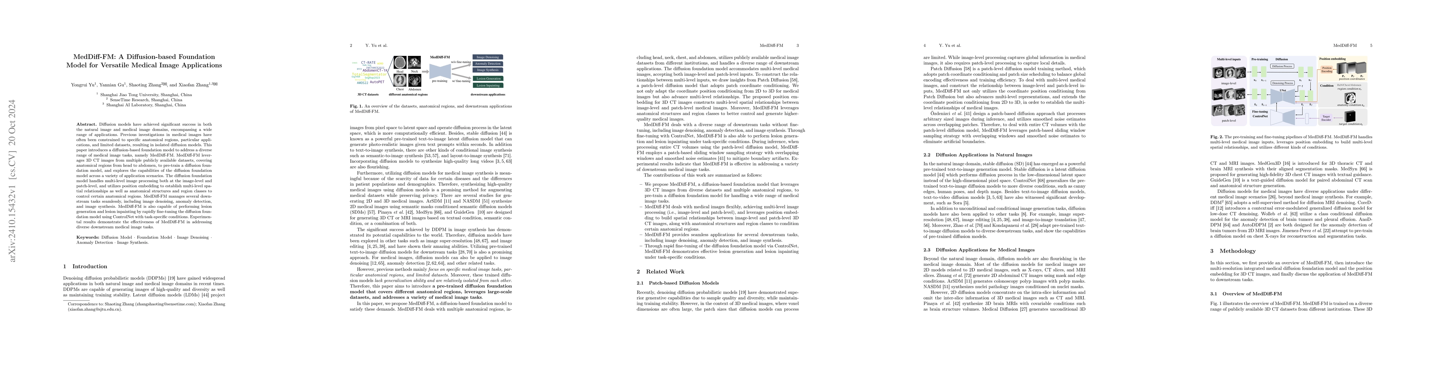
Diffusion models have achieved significant success in both the natural image and medical image domains, encompassing a wide range of applications. Previous investigations in medical images have often been constrained to specific anatomical regions, particular applications, and limited datasets, resulting in isolated diffusion models. This paper introduces a diffusion-based foundation model to address a diverse range of medical image tasks, namely MedDiff-FM. MedDiff-FM leverages 3D CT images from multiple publicly available datasets, covering anatomical regions from head to abdomen, to pre-train a diffusion foundation model, and explores the capabilities of the diffusion foundation model across a variety of application scenarios. The diffusion foundation model handles multi-level image processing both at the image-level and patch-level, and utilizes position embedding to establish multi-level spatial relationships as well as anatomical structures and region classes to control certain anatomical regions. MedDiff-FM manages several downstream tasks seamlessly, including image denoising, anomaly detection, and image synthesis. MedDiff-FM is also capable of performing lesion generation and lesion inpainting by rapidly fine-tuning the diffusion foundation model using ControlNet with task-specific conditions. Experimental results demonstrate the effectiveness of MedDiff-FM in addressing diverse downstream medical image tasks.
AI Key Findings
Get AI-generated insights about this paper's methodology, results, significance, and more — seven facets brought into focus.
Impact
Authors
PDF Preview
Citation Network
Current paper (gray), citations (green), references (blue)
Display is limited for performance on very large graphs.
Discussion 0