Metabolic light absorption, scattering and emission (MetaLASE) microscopy
Publication
Metrics
AI Quick Summary
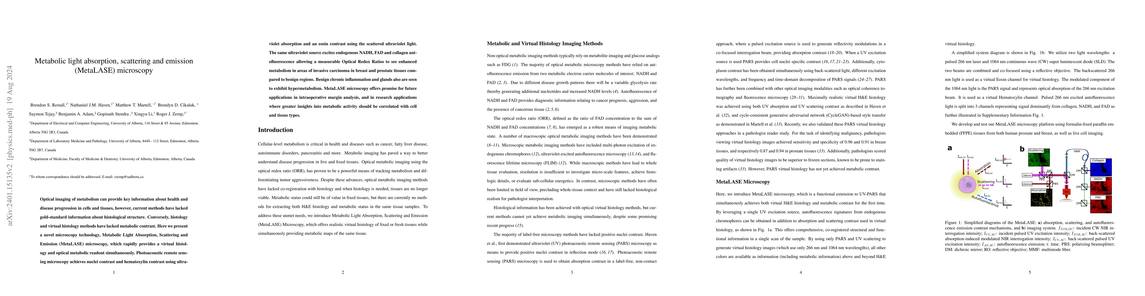
MetaLASE microscopy integrates virtual histology and metabolic imaging, providing simultaneous histological and metabolic contrast, and revealing enhanced metabolism in invasive carcinomas and benign regions. This novel technique holds potential for intraoperative margin analysis and research correlating metabolic activity with tissue types.
Paper Preview
Abstract
Optical imaging of metabolism can provide key information about health and disease progression in cells and tissues, however, current methods have lacked gold-standard information about histological structure. Conversely, histology and virtual histology methods have lacked metabolic contrast. Here we present a novel microscopy technology, Metabolic Light Absorption, Scattering and Emission (MetaLASE) microscopy, which rapidly provides a virtual histology and optical metabolic readout simultaneously. Photoacoustic remote sensing microscopy achieves nuclei contrast and hematoxylin contrast using ultraviolet absorption and an eosin contrast using the scattered ultraviolet light. The same ultraviolet source excites endogenous NADH, FAD and collagen autofluorescence allowing a measurable Optical Redox Ratios to see enhanced metabolism in areas of invasive carcinoma in breast and prostate tissues compared to benign regions. Benign chronic inflammation and glands also are seen to exhibit hypermetabolism. MetaLASE microscopy offers promise for future applications in intraoperative margin analysis, and in research applications where greater insights into metabolic activity should be correlated with cell and tissue types.
AI Key Findings
Get AI-generated insights about this paper's methodology, results, significance, and more — seven facets brought into focus.
Impact
Paper Details
Authors
PDF Preview
Key Terms
Citation Network
Current paper (gray), citations (green), references (blue)
Display is limited for performance on very large graphs.
Discussion 0