Metabolic Reactions Studied by Zero- and Low-Field Nuclear Magnetic Resonance
Publication
Metrics
AI Quick Summary
Researchers successfully used zero- and low-field nuclear magnetic resonance to track metabolic reactions without requiring large superconducting magnets. This breakthrough enables new biomedical imaging applications.
Paper Preview
Abstract
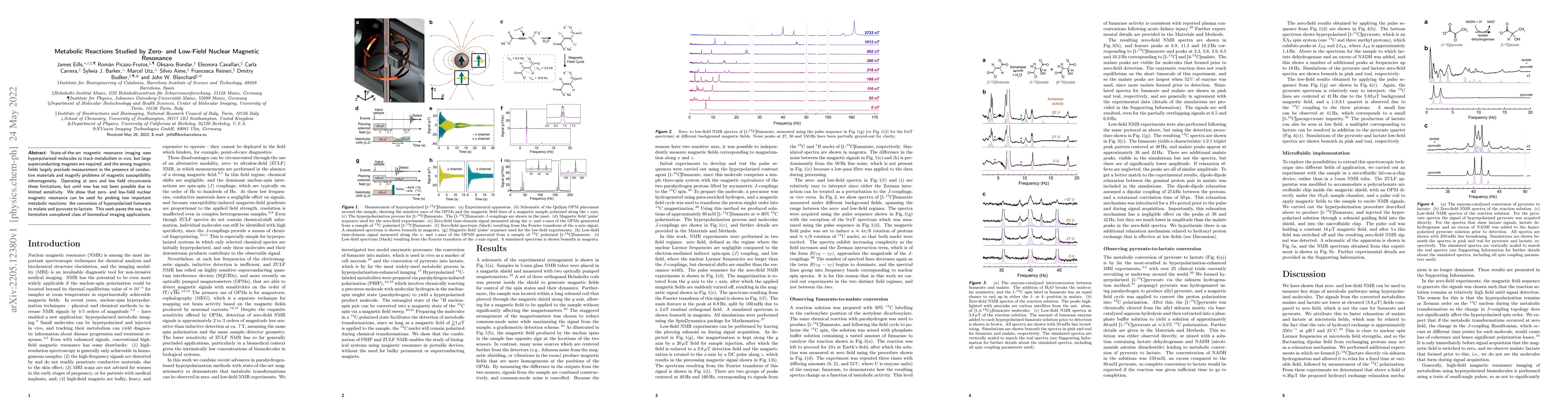
State-of-the-art magnetic resonance imaging uses hyperpolarized molecules to track metabolism in vivo, but large superconducting magnets are required, and the strong magnetic fields largely preclude measurement in the presence of conductive materials and magnify problems of magnetic susceptibility inhomogeneity. Operating at zero and low field circumvents these limitations, but until now has not been possible due to limited sensitivity. We show that zero- and low-field nuclear magnetic resonance can be used for probing two important metabolic reactions: the conversion of hyperpolarized fumarate to malate and pyruvate to lactate. This work paves the way to a heretofore unexplored class of biomedical imaging applications.
AI Key Findings
Get AI-generated insights about this paper's methodology, results, significance, and more — seven facets brought into focus.
Impact
Paper Details
Authors
PDF Preview
Key Terms
Citation Network
Current paper (gray), citations (green), references (blue)
Display is limited for performance on very large graphs.
Discussion 0