AI Quick Summary
This paper proposes a micro-modulated luminescence tomography (MLT) method to enhance contrast and resolution in deep biological imaging by using LiGa5O8:Cr3+ nanophosphors. Numerical simulations show the potential of MLT for high-resolution quantification of nanophosphor distribution in thick samples.
Quick Answers
What methodology did the authors use?
The research proposes micro-modulated luminescence tomography (MLT) to quantify nanophosphor distribution in thick biological samples with high resolution. It involves a micro-focus x-ray source, an x-ray zone plate, two EMCCD cameras, NIR laser stimulation sources, and a rotating stage integrated on an optical table. More in Methodology →
What are the key results?
Numerical simulations demonstrate the feasibility of MLT for nanophosphor distribution quantification in biological samples. — MLT can achieve micron resolution imaging deep into tissue samples due to deep penetration of well-modulated x-ray excitation and NIR laser stimulation of stored luminescence energy. More in Key Results →
Why is this work significant?
MLT is significant for investigating biological features and processes, monitoring drug delivery, assessing cancer therapy, and other applications, overcoming the imaging depth limits of conventional optical microscopic methods. More in Significance →
What are the main limitations?
MLT may require more imaging time compared to other methods like x-ray luminescence tomography and stored luminescence tomography due to its targeted high resolution. — The approach relies on the successful application of an x-ray zone plate, which, although proven in nano-x-ray computed tomography, may present manufacturing challenges at micro-millimeter precision. More in Limitations →
Abstract
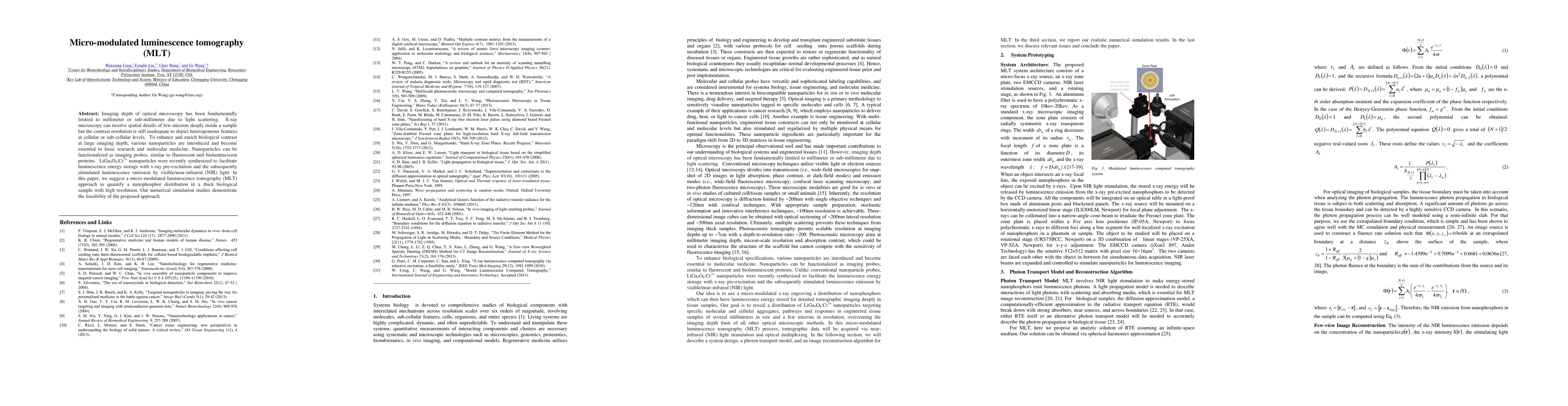
Imaging depth of optical microscopy has been fundamentally limited to millimeter or sub-millimeter due to light scattering. X-ray microscopy can resolve spatial details of few microns deeply inside a sample but the contrast resolution is still inadequate to depict heterogeneous features at cellular or sub-cellular levels. To enhance and enrich biological contrast at large imaging depth, various nanoparticles are introduced and become essential to basic research and molecular medicine. Nanoparticles can be functionalized as imaging probes, similar to fluorescent and bioluminescent proteins. LiGa5O8:Cr3+ nanoparticles were recently synthesized to facilitate luminescence energy storage with x-ray pre-excitation and the subsequently stimulated luminescence emission by visible/near-infrared (NIR) light. In this paper, we suggest a micro-modulated luminescence tomography (MLT) approach to quantify a nanophosphor distribution in a thick biological sample with high resolution. Our numerical simulation studies demonstrate the feasibility of the proposed approach.
AI Key Findings
Generated Sep 07, 2025
Methodology — What approach did the authors take?
The research proposes micro-modulated luminescence tomography (MLT) to quantify nanophosphor distribution in thick biological samples with high resolution. It involves a micro-focus x-ray source, an x-ray zone plate, two EMCCD cameras, NIR laser stimulation sources, and a rotating stage integrated on an optical table.
Key Results — What are the main findings?
- Numerical simulations demonstrate the feasibility of MLT for nanophosphor distribution quantification in biological samples.
- MLT can achieve micron resolution imaging deep into tissue samples due to deep penetration of well-modulated x-ray excitation and NIR laser stimulation of stored luminescence energy.
Significance — Why does this research matter?
MLT is significant for investigating biological features and processes, monitoring drug delivery, assessing cancer therapy, and other applications, overcoming the imaging depth limits of conventional optical microscopic methods.
Technical Contribution — What is the technical contribution?
The main technical contribution of this research is the development of an MLT system architecture and photon transport model for high-resolution nanophosphor distribution imaging in thick biological samples using micro-modulated x-ray engraving and NIR light stimulation.
Novelty — What is new about this work?
MLT distinguishes itself by utilizing LiGa5O8:Cr3+ nanoparticles for luminescence energy storage with x-ray pre-excitation and subsequent NIR light stimulation, enabling detailed tomographic imaging deeply in tissue samples with micron resolution.
Limitations — What are the limitations of this study?
- MLT may require more imaging time compared to other methods like x-ray luminescence tomography and stored luminescence tomography due to its targeted high resolution.
- The approach relies on the successful application of an x-ray zone plate, which, although proven in nano-x-ray computed tomography, may present manufacturing challenges at micro-millimeter precision.
Future Work — What did the authors propose for future work?
- Exploration of few-view tomography techniques to reduce projection views and lower radiation dose in MLT.
- Investigating the utility of MLT in various biological contexts, such as cancer research and tissue engineering.
Paper Details
How to Cite This Paper
@article{anon2015micro,
title = {Micro-modulated luminescence tomography},
year = {2015},
eprint = {1311.4107},
archivePrefix = {arXiv},
primaryClass = {physics.bio-ph},
doi = {10.1364/OE.22.005572},
}
(2015). Micro-modulated luminescence tomography. arXiv. https://doi.org/10.1364/OE.22.005572
"Micro-modulated luminescence tomography." arXiv, 2015, doi.org/10.1364/OE.22.005572.
PDF Preview
Key Terms
Citation Network
Current paper (gray), citations (green), references (blue)
Display is limited for performance on very large graphs.
Similar Papers
Found 4 papers| Title | Authors | Year | Actions |
|---|
Comments (0)