Microdosimetry in ion-beam therapy: studying and comparing outcomes from different detectors
Publication
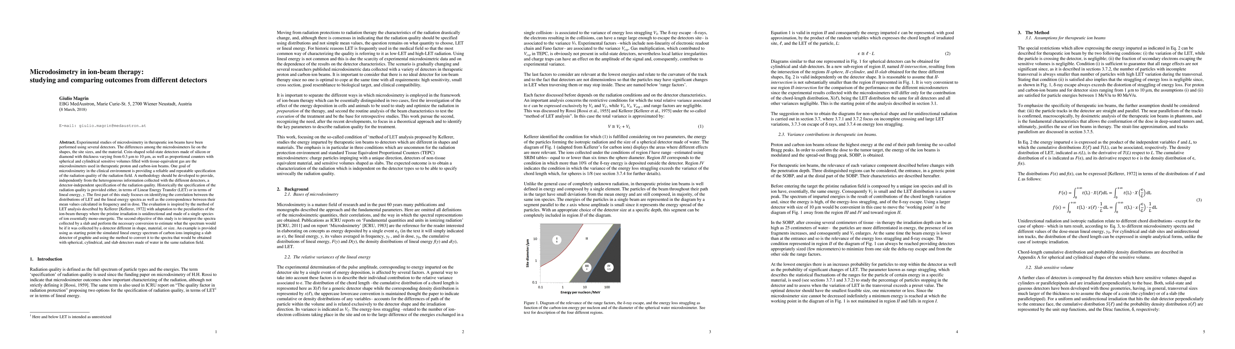
Metrics
Paper Preview
Abstract
Experimental studies of microdosimetry in therapeutic ion beams have been performed using several detectors. The differences among them lie on the shapes, the site sizes, and the material. Coin-shaped solid-state detectors made of silicon or diamond with thickness varying from 0.3 to 10 microns, as well as proportional counters with spherical and cylindrical sensitive volumes filled with tissue-equivalent gas are the microdosimeters used in therapeutic proton and carbon-ion beams. One goal of microdosimetry in the clinical environment is providing repeatable specification of the radiation quality of the radiation field. A methodology should be developed to provide, independently from the heterogeneous information collected with the different detectors, a detector-independent specification of the radiation quality. Historically the specification of the radiation quality is provided either, in terms of Linear Energy Transfer (LET) or in terms of lineal energy, y. First this study focuses on identifying the correlation between the distributions of LET and the lineal energy spectra as well as the correspondence between their mean values calculated in frequency and in dose. The evaluation is based on the method of LET analysis described by Kellerer making the adaptation to the peculiarities of the therapeutic ion-beam where the pristine irradiation is unidirectional and made of a single type of mono-energetic ions. The second objective of this study is to interpret the spectrum collected by a slab and estimate what the spectrum would be if it was collected by a detector different in shape, material, or size. An example confirms the method starting from the simulated lineal energy spectrum obtained for carbon ions in a slab detector of graphite and converting it to the spectruma that would be obtained in the same radiation field for spherical, cylindrical, and slab detector made of water.
AI Key Findings
Get AI-generated insights about this paper's methodology, results, significance, and more — seven facets brought into focus.
Impact
Paper Details
PDF Preview
Key Terms
Citation Network
Current paper (gray), citations (green), references (blue)
Display is limited for performance on very large graphs.
Discussion 0