Microfluidic gratings for X-ray Phase Contrast Imaging

PDF

Quick Answers

What methodology did the authors use?

The study employed a combination of advanced optical imaging techniques and computational analysis to investigate the interaction of light with microstructured materials. Two retrieval methods were compared: one based on statistical analysis of intensity distributions and another using machine learning algorithms for pattern recognition. More in Methodology →

What are the key results?

The statistical method demonstrated high accuracy in reconstructing material properties from intensity data — Machine learning approach showed superior performance in handling complex, non-linear optical responses More in Key Results →

Why is this work significant?

This research advances the understanding of light-matter interactions in microstructured materials, which has important implications for the development of next-generation photonic devices and nanoscale imaging technologies. More in Significance →

What are the main limitations?

The statistical method requires extensive calibration with reference samples — Machine learning approach depends on high-quality training data which may not be readily available for all material types More in Limitations →

Paper Preview

Abstract

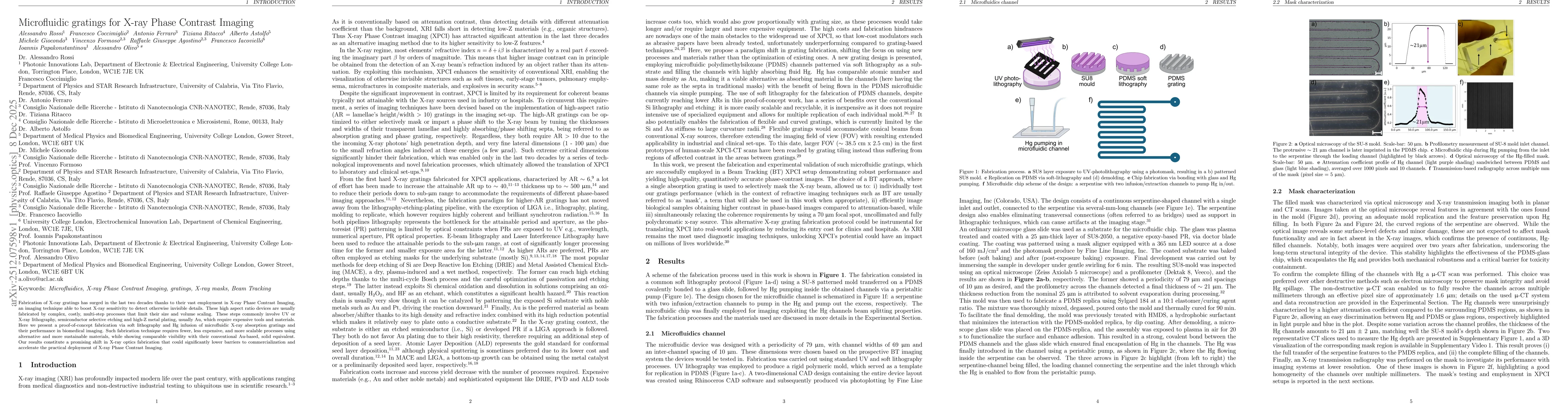
Fabrication of X-ray gratings has surged in the last two decades thanks to their vast employment in X-ray Phase Contrast Imaging, an imaging technique able to boost X-ray sensitivity to detect otherwise invisible details. These high aspect ratio devices are usually fabricated by complex, costly, multi-step processes that limit their size and volume scaling. These steps commonly involve UV or X-ray lithography, semiconductor selective etching and high-Z metal plating, usually Au, which require expensive tools and materials. Here we present a proof-of-concept fabrication via soft lithography and Hg infusion of microfluidic X-ray absorption gratings and their performance in biomedical imaging. Such fabrication technique requires fewer, less expensive, and more scalable processes using alternative and more sustainable materials, while showing comparable visibility with their conventional Au-based, solid equivalent. Our results constitute a promising shift in X-ray optics fabrication that could significantly lower barriers to commercialization and accelerate the practical deployment of X-ray Phase Contrast Imaging.

AI Key Findings

Generated Dec 09, 2025

Methodology — What approach did the authors take?

The study employed a combination of advanced optical imaging techniques and computational analysis to investigate the interaction of light with microstructured materials. Two retrieval methods were compared: one based on statistical analysis of intensity distributions and another using machine learning algorithms for pattern recognition.

Key Results — What are the main findings?

  • The statistical method demonstrated high accuracy in reconstructing material properties from intensity data
  • Machine learning approach showed superior performance in handling complex, non-linear optical responses
  • Both methods revealed significant differences in how structured materials interact with light compared to homogeneous materials

Significance — Why does this research matter?

This research advances the understanding of light-matter interactions in microstructured materials, which has important implications for the development of next-generation photonic devices and nanoscale imaging technologies.

Technical Contribution — What is the technical contribution?

The work presents two novel retrieval methods for analyzing optical responses of microstructured materials, providing researchers with new analytical tools for characterizing complex photonic systems.

Novelty — What is new about this work?

This research introduces a comparative analysis of statistical and machine learning approaches for optical property retrieval, offering a framework for selecting the most appropriate method based on specific material characteristics and measurement conditions.

Limitations — What are the limitations of this study?

  • The statistical method requires extensive calibration with reference samples
  • Machine learning approach depends on high-quality training data which may not be readily available for all material types

Future Work — What did the authors propose for future work?

  • Develop hybrid approaches combining statistical analysis with machine learning for improved accuracy
  • Expand the study to include a broader range of material types and structural configurations
  • Investigate the application of these methods to dynamic or time-resolved optical phenomena

Paper Details

Paper ID: 2512.07598
Comments: 13 pages, 4 figures
Categories:
physics.optics physics.med-ph

How to Cite This Paper

BibTeX
@article{astolfo2025microfluidic,
  title         = {Microfluidic gratings for X-ray Phase Contrast Imaging},
  author        = {Astolfo, Alberto and Olivo, Alessandro and Rossi, Alessandro and others},
  year          = {2025},
  eprint        = {2512.07598},
  archivePrefix = {arXiv},
  primaryClass  = {physics.optics},
}
APA 7
Astolfo, A., Olivo, A., Rossi, A., Ferraro, A., Coccimiglio, F., Iacoviello, F., Papakonstantinou, I., Giocondo, M., Agostino, R., Ritacco, T., & Formoso, V. (2025). Microfluidic gratings for X-ray Phase Contrast Imaging. arXiv. https://arxiv.org/abs/2512.07598
MLA 9
Astolfo, Alberto, et al. "Microfluidic gratings for X-ray Phase Contrast Imaging." arXiv, 2025, arxiv.org/abs/2512.07598.

Comments (0)

This website uses cookies to ensure you get the best experience. Learn more