Micromagnetism in (001) magnetite by spin-polarized low-energy electron microscopy
Publication
Metrics
AI Quick Summary
Researchers used spin-polarized low-energy electron microscopy to map magnetization vectors in a magnetite crystal, finding domains aligned along surface directions and unusual curved domain walls. The study provides new insights into the magnetic structure of magnetite.
Paper Preview
Abstract
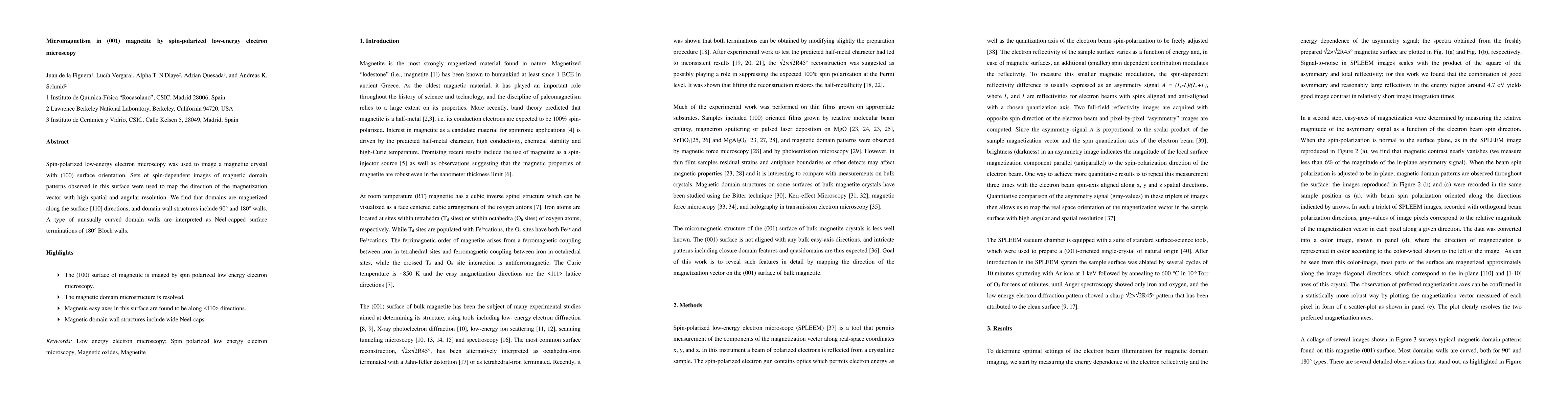
Spin-polarized low-energy electron microscopy was used to image a magnetite crystal with (100) surface orientation. Sets of spin-dependent images of magnetic domain patterns observed in this surface were used to map the direction of the magnetization vector with high spatial and angular resolution. We find that domains are magnetized along the surface [110] directions, and domain wall structures include 90{\deg} and 180{\deg} walls. A type of unusually curved domain walls are interpreted as N\'eel-capped surface terminations of 180{\deg} Bloch walls.
AI Key Findings
Get AI-generated insights about this paper's methodology, results, significance, and more — seven facets brought into focus.
Impact
Paper Details
PDF Preview
Key Terms
Citation Network
Current paper (gray), citations (green), references (blue)
Display is limited for performance on very large graphs.
Discussion 0