Summary

Osteoarthritis (OA) is a multifaceted joint disease which poses significant socioeconomic burdens and remains a significant clinical challenge. Evidence suggests that structural and mechanical changes in subchondral bone influence the pathogenesis and development of OA, leading to diminished bone quality and cartilage degeneration. While changes in microstructure and tissue scale elastic properties are well reported, the tissue yield response of subchondral bone in OA and their correlation with compositional changes have not been investigated. Here, we performed quasistatic micropillar compression and nanoindentation within the subchondral bone plate and trabeculae of hydrated non-diseased (ND) and OA affected specimens retrieved from the distal tibia in vivo. The micropillars, extracted by laser ablation, exhibited a taper angle which mandated the use of an in silico micropillar compression routine to back-calculate elastic modulus and strength of the bone tissue that comprised each micropillar. Elastic modulus remained unchanged between ND and OA subchondral bone, whereas strength increased from 46.0 MPa to 57.3 MPa in OA subchondral trabecular bone but not in the bone plate. Micropillar matched Raman spectroscopy and quantitative backscattered electron imaging revealed mineralisation is the underlying determinant of elastic modulus and strength at the microscale. By combining micromechanical and tissue compositional analyses, we investigated how the mechanical properties are related and how these properties are affected in subchondral bone by OA. Our results may be of value in the development and optimisation of interventions used to alleviate the socioeconomic burdens associated with this debilitating joint disease.
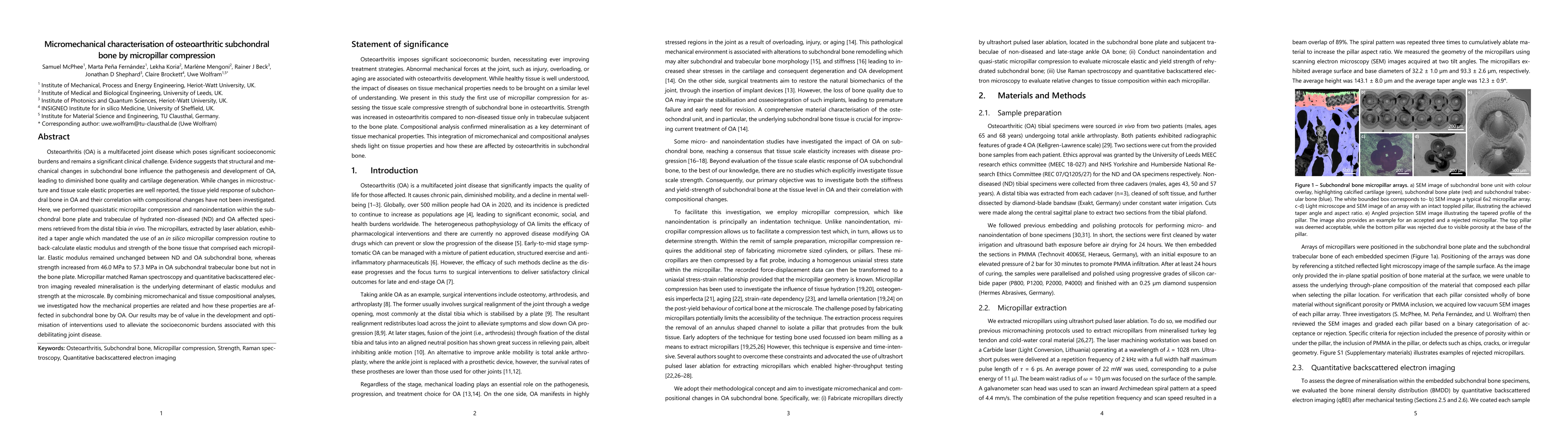
AI Key Findings

Generated Nov 01, 2025

Methodology

The research employed a combination of microscale mechanical testing, polarized Raman spectroscopy, and micropillar compression to analyze the structural and mechanical properties of bone tissue at multiple scales.

Key Results

  • Micropillar compression revealed superior strength and ductility in lamellar bone with no visible damage under high strain rates.
  • Microscale compressive behavior of hydrated bone showed significant strain rate sensitivity, with increased strength at higher strain rates.
  • The integration of polarized Raman spectroscopy with micropillar compression provided insights into the microstructure-property relationships in mineralized tissues.

Significance

This research advances understanding of bone mechanics at the microscale, which is critical for developing better biomaterials and treatments for bone-related diseases and injuries.

Technical Contribution

The study introduced a combined experimental approach using polarized Raman spectroscopy and micropillar compression to study microscale structure-property relationships in mineralized tissues.

Novelty

This work uniquely integrates advanced mechanical testing with spectroscopic analysis to provide comprehensive insights into bone's microstructural and mechanical behavior across multiple scales.

Limitations

  • The study focused primarily on cortical bone and may not fully represent trabecular bone mechanics.
  • Results are based on in vitro experiments, which may not fully capture in vivo physiological conditions.

Future Work

  • Investigate the mechanical behavior of trabecular bone using similar multi-scale techniques.
  • Explore the effects of aging and disease on microscale bone properties.
  • Develop computational models to simulate microscale bone mechanics based on experimental data.

Paper Details

PDF Preview

Download PDF

Comments (0)

This website uses cookies to ensure you get the best experience. Learn more