MicroMIL: Graph-based Contextual Multiple Instance Learning for Patient Diagnosis Using Microscopy Images
Publication
Metrics
AI Quick Summary
MicroMIL presents a graph-based framework for contextual multiple instance learning (MIL) to diagnose patients using microscopy images, addressing issues of unknown positions and redundant captures. It outperforms existing methods by using deep cluster embedding and graph neural networks, achieving better results on colon cancer and BreakHis datasets.
Paper Preview
Abstract
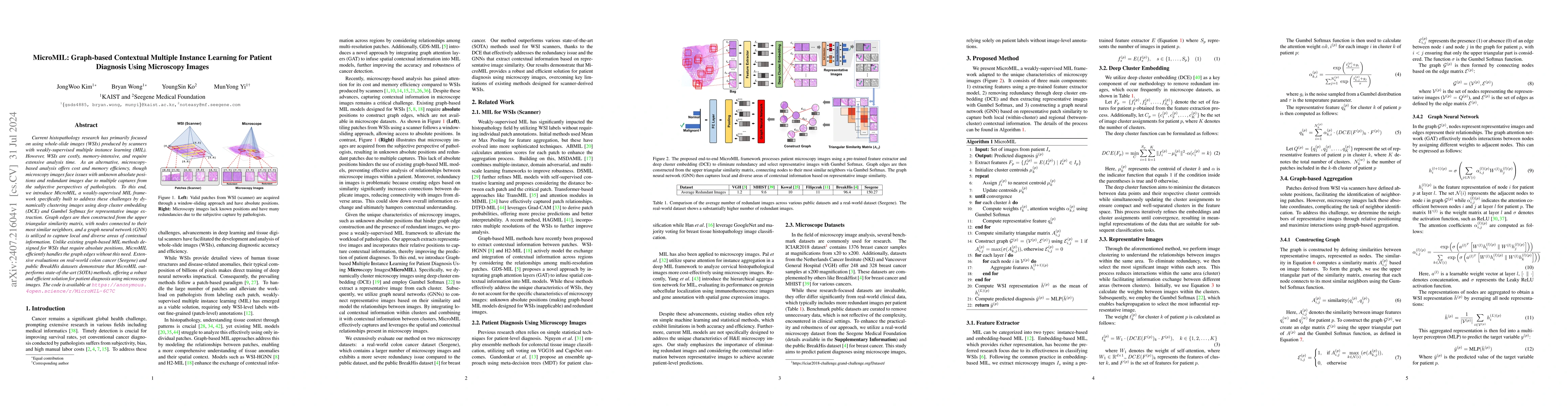
Current histopathology research has primarily focused on using whole-slide images (WSIs) produced by scanners with weakly-supervised multiple instance learning (MIL). However, WSIs are costly, memory-intensive, and require extensive analysis time. As an alternative, microscopy-based analysis offers cost and memory efficiency, though microscopy images face issues with unknown absolute positions and redundant images due to multiple captures from the subjective perspectives of pathologists. To this end, we introduce MicroMIL, a weakly-supervised MIL framework specifically built to address these challenges by dynamically clustering images using deep cluster embedding (DCE) and Gumbel Softmax for representative image extraction. Graph edges are then constructed from the upper triangular similarity matrix, with nodes connected to their most similar neighbors, and a graph neural network (GNN) is utilized to capture local and diverse areas of contextual information. Unlike existing graph-based MIL methods designed for WSIs that require absolute positions, MicroMIL efficiently handles the graph edges without this need. Extensive evaluations on real-world colon cancer (Seegene) and public BreakHis datasets demonstrate that MicroMIL outperforms state-of-the-art (SOTA) methods, offering a robust and efficient solution for patient diagnosis using microscopy images. The code is available at https://anonymous.4open.science/r/MicroMIL-6C7C
AI Key Findings
Get AI-generated insights about this paper's methodology, results, significance, and more — seven facets brought into focus.
Impact
Paper Details
Authors
PDF Preview
Key Terms
Citation Network
Current paper (gray), citations (green), references (blue)
Display is limited for performance on very large graphs.
Discussion 0