Publication
Metrics
Quick Actions
AI Quick Summary
Microscopic Propagator Imaging (MPI) is a novel method for retrieving indices of the microscopic propagator, which reflects water displacements due to diffusion within neural microstructures. Unlike other diffusion MRI techniques, MPI indices are independent of tissue organization, making them more specific to microstructures like axons and cells, and thus more sensitive to their presence and integrity.
Quick Answers
What is "Microscopic Propagator Imaging (MPI) with Diffusion MRI" about?
Microscopic Propagator Imaging (MPI) is a novel method for retrieving indices of the microscopic propagator, which reflects water displacements due to diffusion within neural microstructures. Unlike other diffusion MRI techniques, MPI indices are independent of tissue organization, making them more specific to microstructures like axons and cells, and thus more sensitive to their presence and integrity.
What methodology did the authors use?
The research introduces Microscopic Propagator Imaging (MPI) using Diffusion MRI, which retrieves indices of the microscopic propagator, independent of mesoscopic tissue organization. It employs zonal modeling of spherical harmonics, signal simulation, and machine learning regression, demonstrated on synthetic and human MRI data. More in Methodology →
What are the key results?
MPI indices are specific to microstructure volumes, sizes, and types, allowing direct linkage to alterations in microstructure presence and integrity. — MPI outperforms Ensemble Average Propagator and Diffusion Tensor Imaging metrics by providing indices independent of tissue orientation dispersion. More in Key Results →
Why is this work significant?
This research offers a novel method to quantitatively describe tissue microstructures without bias from mesoscopic orientational organization, potentially enhancing diagnostic relevance and interpretability compared to conventional methods. More in Significance →
What are the main limitations?
Parallel non-Gaussianity (NG∥) was found to be unreliable, possibly due to insufficient simulation of parallel non-Gaussianity for axons in the training dataset. — The study did not address the presence of exchange between tissue compartments in the simulated kernel signal, which may not be straightforward to quantify with the current q-space acquisition scheme. More in Limitations →
Abstract
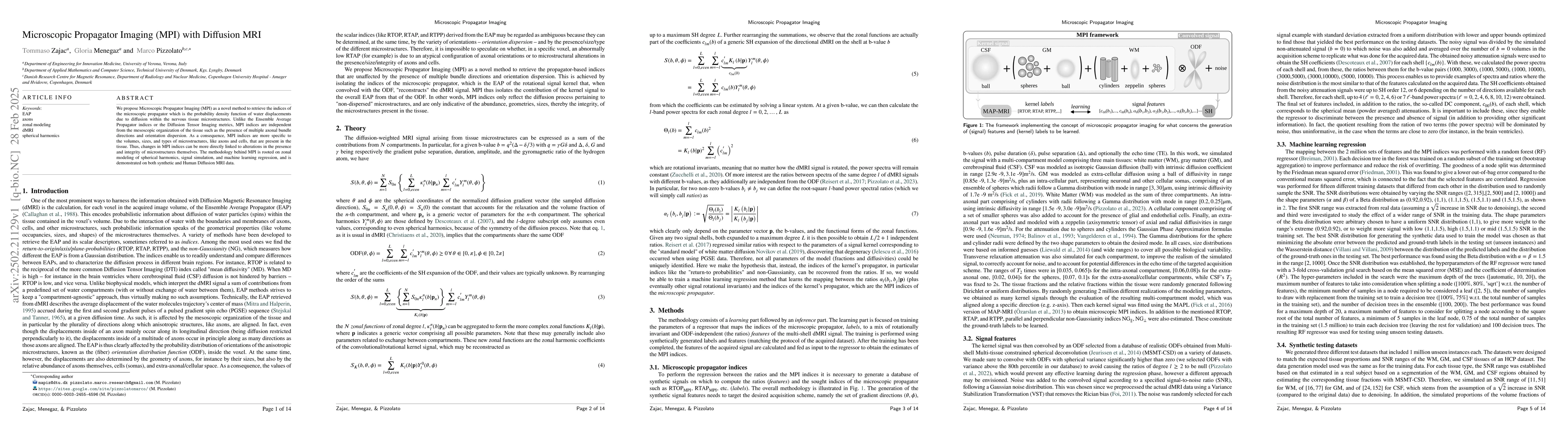
We propose Microscopic Propagator Imaging (MPI) as a novel method to retrieve the indices of the microscopic propagator which is the probability density function of water displacements due to diffusion within the nervous tissue microstructures. Unlike the Ensemble Average Propagator indices or the Diffusion Tensor Imaging metrics, MPI indices are independent from the mesoscopic organization of the tissue such as the presence of multiple axonal bundle directions and orientation dispersion. As a consequence, MPI indices are more specific to the volumes, sizes, and types of microstructures, like axons and cells, that are present in the tissue. Thus, changes in MPI indices can be more directly linked to alterations in the presence and integrity of microstructures themselves. The methodology behind MPI is rooted on zonal modeling of spherical harmonics, signal simulation, and machine learning regression, and is demonstrated on both synthetic and Human Diffusion MRI data.
AI Key Findings
Generated Jun 10, 2025
Methodology — What approach did the authors take?
The research introduces Microscopic Propagator Imaging (MPI) using Diffusion MRI, which retrieves indices of the microscopic propagator, independent of mesoscopic tissue organization. It employs zonal modeling of spherical harmonics, signal simulation, and machine learning regression, demonstrated on synthetic and human MRI data.
Key Results — What are the main findings?
- MPI indices are specific to microstructure volumes, sizes, and types, allowing direct linkage to alterations in microstructure presence and integrity.
- MPI outperforms Ensemble Average Propagator and Diffusion Tensor Imaging metrics by providing indices independent of tissue orientation dispersion.
- A random forest regression model effectively maps between 2 million feature sets and MPI indices, validated across various SNR distributions.
- Synthetic testing datasets accurately mimicked tissue proportions and SNR ranges of WM, GM, and CSF from HCP data.
- MPI indices show good regression performance for microscopic RTOP, RTAP, and RTPP, with narrower error distributions in GM and CSF compared to WM.
Significance — Why does this research matter?
This research offers a novel method to quantitatively describe tissue microstructures without bias from mesoscopic orientational organization, potentially enhancing diagnostic relevance and interpretability compared to conventional methods.
Technical Contribution — What is the technical contribution?
The proposed MPI framework decouples the simulation of the kernel signal from the estimated indices, enabling general and unbiased quantitative description of diffusion processes due to microstructures.
Novelty — What is new about this work?
MPI distinguishes itself by providing ODF-invariant indices related solely to microscopic tissue properties, unlike conventional methods sensitive to mesoscopic orientational organization.
Limitations — What are the limitations of this study?
- Parallel non-Gaussianity (NG∥) was found to be unreliable, possibly due to insufficient simulation of parallel non-Gaussianity for axons in the training dataset.
- The study did not address the presence of exchange between tissue compartments in the simulated kernel signal, which may not be straightforward to quantify with the current q-space acquisition scheme.
Future Work — What did the authors propose for future work?
- Explore more sophisticated synthetic signal generation strategies, such as Monte Carlo simulations, to improve the realism of MPI indices and enable learning of additional microscopic propagator features.
- Investigate the possibility of bypassing the fitting of MAP-MRI to the simulated kernel signal using Monte Carlo simulations, which could calculate indices directly from simulated random walker trajectories.
Paper Details
How to Cite This Paper
@article{menegaz2025microscopic,
title = {Microscopic Propagator Imaging (MPI) with Diffusion MRI},
author = {Menegaz, Gloria and Pizzolato, Marco and Zajac, Tommaso},
year = {2025},
eprint = {2502.21129},
archivePrefix = {arXiv},
primaryClass = {q-bio.NC},
}
Menegaz, G., Pizzolato, M., & Zajac, T. (2025). Microscopic Propagator Imaging (MPI) with Diffusion MRI. arXiv. https://arxiv.org/abs/2502.21129
Menegaz, Gloria, et al. "Microscopic Propagator Imaging (MPI) with Diffusion MRI." arXiv, 2025, arxiv.org/abs/2502.21129.
PDF Preview
Citation Network
Current paper (gray), citations (green), references (blue)
Display is limited for performance on very large graphs.
Similar Papers
Found 4 papersMicroscopic Propagator Imaging with diffusion MRI.
Menegaz, Gloria, Zajac, Tommaso, Pizzolato, Marco
Diffusion MRI experimental design optimization for microstructure imaging.
Chen, Yongxin, Lenglet, Christophe, Farooq, Hamza et al.
Microstructure imaging of prostate cancer by diffusion MRI.
Wu, Dan, Szczepankiewicz, Filip, Palombo, Marco et al.
No citations found for this paper.
Comments (0)