Microscopy Cell Segmentation via Adversarial Neural Networks
Publication
Metrics
AI Quick Summary
A novel cell segmentation method using adversarial neural networks achieves accurate results with minimal annotated data, promising applications in microscopy imaging.
Paper Preview
Abstract
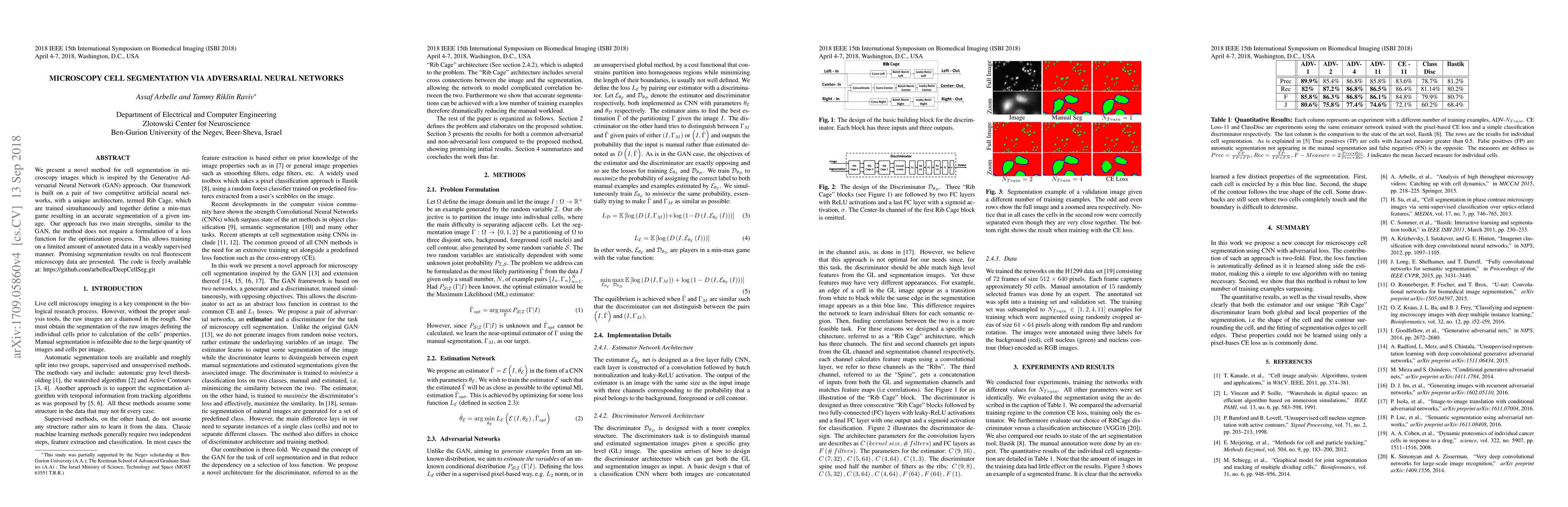
We present a novel method for cell segmentation in microscopy images which is inspired by the Generative Adversarial Neural Network (GAN) approach. Our framework is built on a pair of two competitive artificial neural networks, with a unique architecture, termed Rib Cage, which are trained simultaneously and together define a min-max game resulting in an accurate segmentation of a given image. Our approach has two main strengths, similar to the GAN, the method does not require a formulation of a loss function for the optimization process. This allows training on a limited amount of annotated data in a weakly supervised manner. Promising segmentation results on real fluorescent microscopy data are presented. The code is freely available at: https://github.com/arbellea/DeepCellSeg.git
AI Key Findings
Get AI-generated insights about this paper's methodology, results, significance, and more — seven facets brought into focus.
Impact
Paper Details
PDF Preview
Key Terms
Citation Network
Current paper (gray), citations (green), references (blue)
Display is limited for performance on very large graphs.
Discussion 0