Microscopy Image Segmentation via Point and Shape Regularized Data Synthesis
Publication
Metrics
AI Quick Summary
This paper proposes a method for microscopy image segmentation using point annotations and synthetic data generation, achieving high performance with less labor-intensive data compared to traditional dense annotations. The framework synthesizes training data and trains a segmentation model that outperforms baselines and matches results of models trained on fully annotated images.
Paper Preview
Abstract
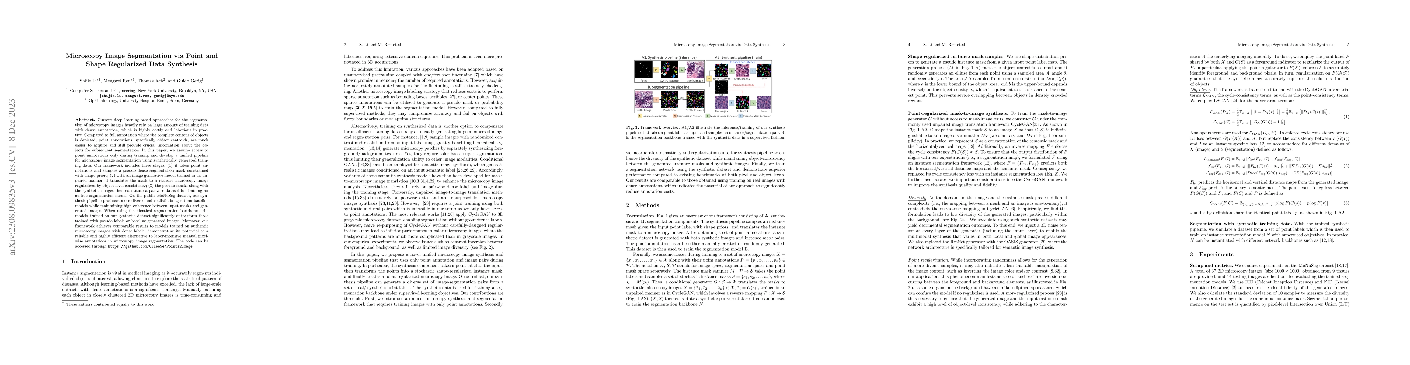
Current deep learning-based approaches for the segmentation of microscopy images heavily rely on large amount of training data with dense annotation, which is highly costly and laborious in practice. Compared to full annotation where the complete contour of objects is depicted, point annotations, specifically object centroids, are much easier to acquire and still provide crucial information about the objects for subsequent segmentation. In this paper, we assume access to point annotations only during training and develop a unified pipeline for microscopy image segmentation using synthetically generated training data. Our framework includes three stages: (1) it takes point annotations and samples a pseudo dense segmentation mask constrained with shape priors; (2) with an image generative model trained in an unpaired manner, it translates the mask to a realistic microscopy image regularized by object level consistency; (3) the pseudo masks along with the synthetic images then constitute a pairwise dataset for training an ad-hoc segmentation model. On the public MoNuSeg dataset, our synthesis pipeline produces more diverse and realistic images than baseline models while maintaining high coherence between input masks and generated images. When using the identical segmentation backbones, the models trained on our synthetic dataset significantly outperform those trained with pseudo-labels or baseline-generated images. Moreover, our framework achieves comparable results to models trained on authentic microscopy images with dense labels, demonstrating its potential as a reliable and highly efficient alternative to labor-intensive manual pixel-wise annotations in microscopy image segmentation. The code is available.
AI Key Findings
Get AI-generated insights about this paper's methodology, results, significance, and more — seven facets brought into focus.
Impact
Paper Details
Authors
PDF Preview
Key Terms
Citation Network
Current paper (gray), citations (green), references (blue)
Display is limited for performance on very large graphs.
Discussion 0