Microscopy of Bioelectric Potentials using Electrochromism
Publication
Metrics
Paper Preview
Abstract
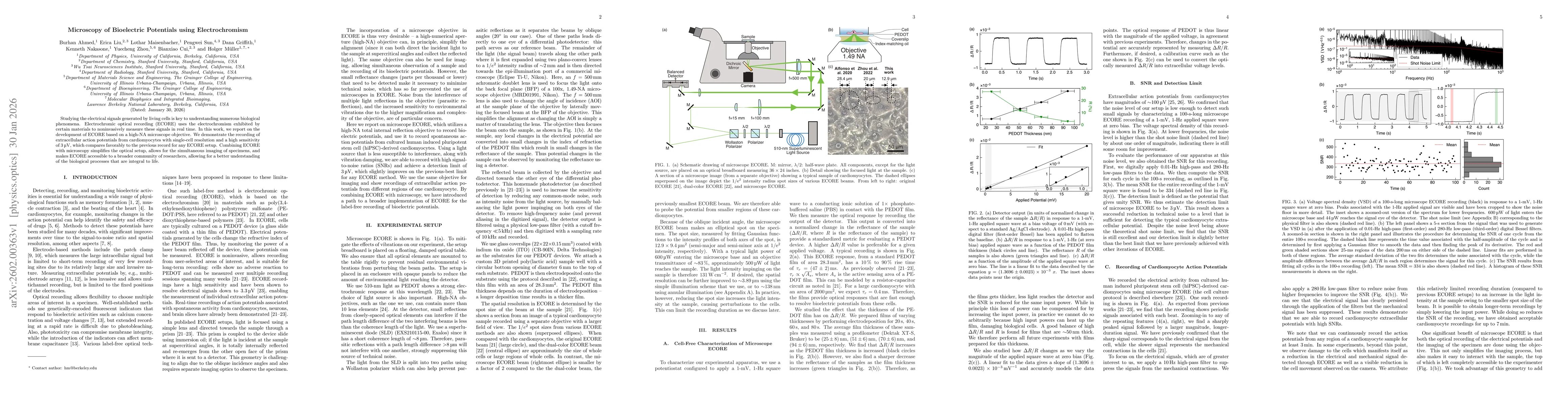
Studying the electrical signals generated by living cells is key to understanding numerous biological phenomena. Electrochromic optical recording (ECORE) uses the electrochromism exhibited by certain materials to noninvasively measure these signals in real time. In this work, we report on the development of ECORE based on a high-NA microscope objective. We demonstrate the recording of extracellular action potentials from cardiomyocytes with single-cell resolution and a high sensitivity of 3 μV, which compares favorably to the previous record for any ECORE setup. Combining ECORE with microscopy simplifies the optical setup, allows for the simultaneous imaging of specimens, and makes ECORE accessible to a broader community of researchers, allowing for a better understanding of the biological processes that are integral to life.
AI Key Findings
Get AI-generated insights about this paper's methodology, results, significance, and more — seven facets brought into focus.
Discussion 0